Editor’s intro: The steps to a full digital workflow include implementing 3D imaging, digital impressions, 3D printed models, and digital practice management — Dr. Thomas Pitts explains the benefits to the doctor, practice, and patient.
Dr. Thomas R. Pitts discusses how digital technology can benefit the doctor, practice, and patients

Many orthodontic practices seeking excellence are moving into the direction of a “fully digital workflow.” What does a fully digital workflow include?
- Going from impression material to scanning, eliminating alginate
- Digital models instead of mounted plaster casts
- Working with a digital lab that will use only 3D-printed models for all appliances made in lab
- Digital smile design for bracket placement
- Digital 3D imaging for radiology and hard tissue measurements
- Three-dimensional volumetric measurements for airway analysis (Figure 3)
- Digital statistics for practice management
- And, of course, digital charting and image collection
It’s a lot to take in. Even when focusing on just one aspect of a digital workflow — imaging in this case — the decision to invest in digital technology is not to be taken lightly. However, ultimately, it’s one that will benefit doctor, practice, and patient alike.
No going back

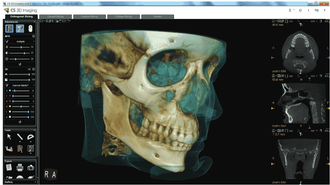
Most orthodontic practices that are committed to excellence have either already made the switch to digital or are seriously considering transitioning to 3D imaging for their patients. Cone beam computed tomography (CBCT) is a great diagnostic and communication tool; it’s become an absolute “must” in my practice for every patient. I use the CS 9300C (Carestream Dental), along with CS 3D Imaging and CS Orthodontic imaging software. In addition to 3D imaging, this system also allows for 2D and necessary cephalometric imaging; or a lateral ceph and a panoramic image can be quickly rendered from the 3D scan (Figure 1). Having used this system for 5 years, there is no way that I would go back to 2D. Not only does CBCT imaging give a more accurate view of the exact positioning of all teeth (Figure 2), roots and erupting teeth, it’s essential for studying the airway; evaluating bone density for mini implants; and understanding the exact relationships of impacted teeth to adjacent teeth.
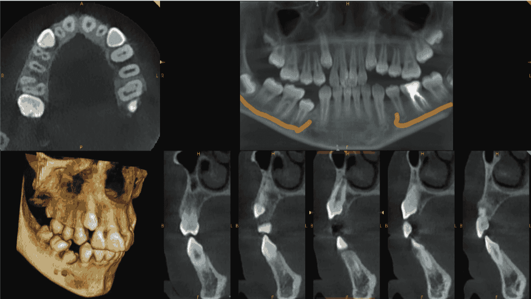
Figure 3: Airway analysis is a growing field in orthodontics (left) and Figure 4: 3D imaging shows the relationship of impacted teeth (right)
Often, CBCT imaging reveals a super-numerary that is hard to see in a traditional 2D panoramic image. In addition to enhancing a doctor’s diagnoses and expanding treatment capabilities, CBCT images and slices are excellent communication tools for patients and parents to follow an eruption path, locate impacted positions, show forces needed to correct inclinations and identify mesiodens and other supernumeraries (Figures 4 and 5). Naturally, these easy-to-view-and-share 3D images also go a long when communicating with a primary care dentist as well.
Next steps in the digital workflow
In addition to my CBCT system, my fully digital workflow includes an intraoral scanner (CS 3600, Carestream Dental), modeling software (CS Model +, Carestream Dental), and CS OrthoTrac practice management software. Now, to move my practice even more toward excellence, the next step to enhancing my fully digital workflow is the addition of a 3D printer in order to print TMD, sleep and intraoral appliances in-house from scanned models. This will save time and money in the future by using past scanned images captured with the intraoral scanner for the fabrication of lost or broken appliances. The patient will need to come in for only the delivery appointment, not an additional impression appointment. This methodology will also speed up the time for delivery of appliances in practices with an in-house lab. The open architecture of my existing digital technology allows me to invest in the printer of my choice.

Introducing digital technology to my practice has made achieving excellence in orthodontics easier than ever. Once you have practiced with the speed and accuracy of digital 3D imaging, there’s no going back to traditional methods.
Besides outlining the steps to a full digital workflow, Dr. Pitts has also shared his expertise with Orthodontic Practice US readers on the subject of increasing consistency using a straight-wire appliance. Read his article here.
Stay Relevant With Orthodontic Practice US
Join our email list for CE courses and webinars, articles and mores

 Tom Pitts, DDS, MSD, graduated from the University of Washington’s orthodontic program in 1970. After graduating, he simultaneously practiced and consulted orthodontists on practice management and clinical protocols. Pitts previously served as a consultant for Ormco Corp, in Anaheim, California, and OC Orthodontics in Oregon. He was an associate clinical professor in orthodontics at the University of the Pacific School of Dentistry for 20 years and now teaches at the University of Las Vegas orthodontic program. Pitts currently resides in Reno, Nevada, and practices orthodontics with his son. He travels the world presenting lectures and training in clinical esthetic orthodontics.
