Dr. Bob Philpott looks at the developments in adhesive systems and endodontics
The use of composite in the restoration of root-filled teeth has become more common, with the need for esthetic dentistry in general increasing due to patient demands, the increased simplicity of the available systems, and the marketing of the products. Endodontically treated teeth, however, pose different problems. They are often badly broken down with little residual dentin remaining; have altered biological and chemical properties (Sim, et al., 2008; Grigoratos, et al., 2001) due to the effects of treatment procedures on them (Reeh, et al., 1989); and a question has always remained regarding the ability to bond to the altered dentin on the pulp chamber floor and within the root canal (Schüpbach, Krejci, Lutz, 1997; Ferrari, et al., 2000). The predominance of in vitro over in vivo studies on the topic has done little to assist in addressing these concerns.
Studies have shown that the quality of the final coronal restoration is a prognostic factor in the outcome of root canal treatment (Ray, Trope, 1995; Hommez, Coppens, De Moor, 2002; Ng, Mann, Gulabivala, 2011). Studies have also previously compared various materials, their placement (direct versus indirect), microleakage, and performance over time. In the case of composite, concerns have been raised especially about the hydrolytic degradation of the dentin bond over time (Söderholm, et al., 1984; Drummond, 2008).
Placement of composite restorations is technique-sensitive, involving a number of interconnected steps. Studies have shown that adequate isolation, a three-step bonding technique (Foxton, Mannocci, Melo, 2006), incremental placement, and correct finishing are all important in the optimization of outcomes.
 In the case of endodontically treated teeth, there are additional concerns — the altered structure both physically and chemically, the need to protect the root filling, and the performance of direct restorations in badly broken down teeth over time. There are also further technical issues to be considered:
- What is the protocol to clean the access cavity following obturation?
- What is the best material to seal the canal orifices?
- What matrix system is best to aid placement?
- How do direct composites perform in comparison to indirect cuspal coverage restorations?
- What role do adhesive post systems have to play?
Preparation of the access cavity
Depending on the technique used, there are varying amounts of obturation material in the access cavity on completion. Studies have shown that residual eugenol can have a negative effect on the polymerization of composite resin (Millstein, Nathanson, 1983).
Meticulous cleaning of the access cavity is essential. Remnants of root filling material must first be removed using either an ultrasonic scaler or an excavator. The cavity walls should be wiped clean using a cotton pellet soaked in chloroform, the most effective solvent of gutta percha available. The cavity should then be cleaned using a cotton pellet soaked in isopropyl alcohol. This should remove all visible remnants of obturation material, and although concerns have been expressed about eugenol residues remaining in the dentinal tubules, this does not appear to be clinically significant.
Three-step bonding techniques appear to offer better outcomes. The protocol used in the cases below involved etching with 37% phosphoric acid for 20 seconds followed by washing and drying (with care taken to avoid dessication of dentin). Following application of primer, the bonding agent is applied in a thin layer and cured. It is important to avoid pooling of the bonding agent by applying compressed air due to the effects it may have on the final restoration margins.
Sealing of the root canal orifices
A number of materials have been used to seal the root canal orifices following obturation. These include amalgam, glass ionomer cement, IRM, and other zinc oxide and eugenol-based cements, Cavit™ (3M™ ESPE™), and various types of composite resin (Belli, et al., 2001; Galvan, et al., 2002).
Unfortunately, leakage studies have many drawbacks and do not accurately represent the clinical scenario. Comparisons between the various materials have also been inconclusive, with a variety of materials all shown to be effective barriers. Another critical factor in relation to this stage of treatment is the ease of placement. Difficulties are often encountered in the placement of a material like GIC into the coronal portion of the obturated root canal. Incorrect placement of the material can often result in a void between the obturation material and the orifice sealer with potential implications for the prognosis of the treatment.
Concerns have previously been voiced about the use of a flowable composite to ircumvent this problem. Studies have shown that these materials are as effective (Belli, et al., 2001) as many of the alternatives, and due to their flow properties, correct placement becomes less technique-sensitive.
Often, longer 23g and 25g hypodermic needle tips can be used in order to aid visibility during placement when working within the confines of the coronal portions of the canal and pulp chamber. Newer materials such as Dentsply Smart Dentin Replacement (SDR®) have simplified the process even further. This material’s flow characteristics allow it to spread into the available space due to its self-leveling capability. The material has also been marketed as a bulk fill flowable resin, with the option to place it in increments of up to 4 mm due to decreased shrinkage stresses during polymerization (Ilie, Hickel, 2011). These two special properties are especially practical in the restoration of the root-filled tooth in dealing with the anatomical irregularities of the pulp chamber and the necessity for large amounts of restorative material (Figures 1A and 1B).
Matrix systems
Systems such as AutoMatrix® (Dentsply) have been used effectively for many years, especially in the placement of amalgam restorations in root-filled teeth. The importance of the coronal restoration in the outcome of endodontic treatment has been established although this relates primarily to the prevention of ingress of bacteria toward the root filling (Ng, Mann, Gulabivala, 2011).
The importance of establishing a correct and cleansable contact area between root-filled and adjacent teeth holds particular significance when placing direct cuspal coverage restorations. Again, root-filled teeth pose particular problems. The lack of buccal and/or lingual walls often precludes correct placement of a non-sectional matrix, ultimately resulting in large open contact areas. The patient’s inability to maintain correct oral hygiene measures then has obvious implications for the periodontal health of the region. Secondly, depending on the extent of the previous restoration or caries, clinicians may often encounter deep mesial or distal proximal boxes.
Sectional matrices, such as the Palodent® and Palodent® Plus systems (Dentsply), have simplified the process. These systems consist of pre-curved metal strips of varying width and design, a metal V-ring to adapt the margins of the band to tooth tissue, and easy placement wedges of various sizes.
Direct versus indirect restorations
Much of the endodontic literature has focused on the advantages of placement of indirect cuspal coverage restorations on root-filled teeth. In 2002, Aquilino and Caplan concluded that endodontically treated teeth not crowned after treatment were lost at a 6 times greater rate.
Tickle, et al. (2008), in a study on the outcome of NHS endodontic treatment, agreed that placement of a cast cuspal coverage restoration had a positive effect on outcome, although success criteria were not stringent. The basis of this is due to the fact that endodontically treated teeth are often badly broken down, have altered properties due to treatment processes, and have lost their proprioceptive capability (Reeh, Messer, Douglas, 1989). As a result, these teeth appear to be more susceptible to catastrophic fracture and benefit greatly from coverage of the remaining unsupported cusps.
The reality is, however, that cuspal coverage can also be provided directly. Studies comparing both approaches have shown that they are comparable in terms of longevity (Mannocci, Ferrari, Watson, 2001; Mannocci, et al., 2002). More regular follow-up of such cases may be necessary if they are used as long-term restorations, and a question may remain over the effects of saliva on the dentin bond over time.
In the current economic climate, these direct multi-surface composite and amalgam restorations afford both the patient and the clinician the luxury of a tooth protected from fracture, thereby allowing a period of review to establish healing prior to possible placement of a crown. This may be particularly relevant in a tooth of questionable prognosis.
Access through existing cast restorations is also a common scenario encountered during endodontic treatment, assuming satisfactory margins both clinically and radiographically. The protocol for composite restoration placement in these teeth is different.

Figures 1A and 1B: Access cavities following cleaning using outlined protocol prior to restoration placement. Note orifices sealed with IRM
Figures 2A and 2B: Restoration of tooth UL6 using adhesive gold onlay (note cementation under rubber dam)
Longer etching times using hydrofluoric acid and bonding techniques incorporating silane in the protocol are essential in the optimization of outcomes.
Posts: yes or no?
The decision on whether a post is necessary in the restoration of a root-filled tooth is often a difficult one. Restorability of teeth, despite various attempts to quantify it, is a subjective concept (McDonald, Setchell, 2005; Bandlish, McDonald, Setchell, 2006). It has been established that the longevity of such restorations is dependent on the presence of a ferrule of remaining dentin being present. Various figures have been proposed and a minimum of 1-1.5 mm of remaining supragingival tooth tissue seems to be necessary in achieving optimal outcomes.
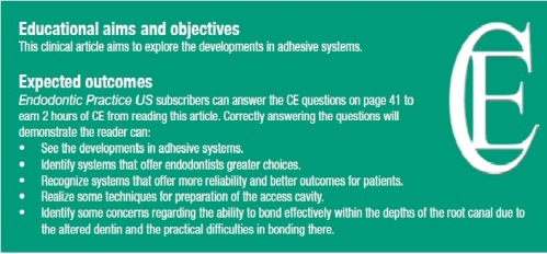
Traditionally, metal (both direct and in-direct) post-and-core systems have been more popular and have been shown to perform well (Nanayakkara, McDonald, Setchell, 1999). Concerns have been raised about the esthetics, removal of tooth tissue, stress effects on remaining dentin (in the case of threaded active posts), and performance in function.More recently, esthetic post systems have become popular, citing improvements in esthetics, ease of manipulation, and functional advantages over metal systems due to their similar modulus of elasticity to dentin. Many studies, both in vitro and in vivo, have concluded that the systems are comparable (Mannocci, et al., 2002) while a systematic review on the subject (Theodosopoulou, Chochlidakis, 2009) concluded that carbon fiber posts in resin matrices appeared to outperform precious metal alloy dowels. Glass fiber posts also appeared to perform better than their metal screw counterparts. Concerns have been raised over the ability to bond effectively within the depths of the root canal due to the altered dentin and the practical difficulties in bonding there. Care must be taken to follow the correct cleaning protocol as outlined, and evidence seems to support the use of dual-cure adhesives in these cases. It has also been suggested that application of hydrogen peroxide and silane to the posts may increase interfacial strengths of such systems in the root canal (Monticelli, et al., 2006). In following these guidelines, esthetic post systems may be particularly beneficial in the restoration of immature anterior teeth, offering the opportunity to reinforce the weakened cervical area.
Conclusion
Developments in adhesive systems have simplified the life of the restorative dentist in recent years, with greater choice and more reliable systems leading to better outcomes for patients. Despite this, the same principles apply and composite restoration placement still remains a technique-sensitive process. Great care must be taken to adhere to strict technical and biological principles in its use.
References
1. Aquilino SA, Caplan DJ. Relationship between crown placement and the survival of endodontically treated teeth. J Prosthet Dent. 2002;87(3):256-263.
2. Bandlish RB1, McDonald AV, Setchell DJ. Assessment of the amount of remaining coronal dentine in root-treated teeth. J Dent. 2006;34(9):699-708.
3. Belli S, Zhang Y, Pereira PN, Ozer F, Pashley DH. Regional bond strengths of adhesive resins to pulp chamber dentin. J Endod. 2001;27(8):527-532.
4. Drummond JL. Degradation, fatigue, and failure of resin dental composite materials. J Dent Res. 2008;87(8):710-719.
5. Ferrari M, Mannocci F, Vichi A, Goracci G. Bond strengths of a porcelain material to different abutment substrates. Oper Dent. 2000;25(4):299-305.
6. Foxton RM, Mannocci F, Melo L. Adhesive restoration of endodontically treated teeth-current research. Dent Update. 2006;33(8):500-502, 505-506.
7. Galvan RR Jr, West LA, Liewehr FR, Pashley DH. Coronal microleakage of five materials used to create an intracoronal seal in endodontically treated teeth. J Endod. 2002;28(2):59-61.
8. Grigoratos D, Knowles J, Ng YL, Gulabivala K. Effect of exposing dentine to sodium hypochlorite and calcium hydroxide on its flexural strength and elastic modulus. Int Endod J. 2001;34(2):113-119.
9. Hommez GM, Coppens CR, De Moor RJ. Periapical health related to the quality of coronal restorations and root fillings. Int Endod J. 2002;35(8):680-689.
10. Ilie N, Hickel R. Investigations on a methacrylate-based flowable composite based on the SDR™ technology. Dent Mater. 2011;27(4):348-355
11. Mannocci F, Ferrari M, Watson TF. Microleakage of endodontically treated teeth restored with fiber posts and composite cores after cyclic loading: a confocal microscopic study. J Prosthet Dent. 2001;85(3):284-291.
12. Mannocci F, Bertelli E, Sherriff M, Watson TF, Ford TR. Three-year clinical comparison of survival of endodontically treated teeth restored with either full cast coverage or with direct composite restoration. J Prosthed Dent. 2002;88(3):297-301.
13. McDonald A, Setchell D. Developing a tooth restorability index. Dent Update. 2005;32(6):343-344, 346-348.
14. Millstein PL, Nathanson D. Effect of eugenol and eugenol cements on cured composite resin. J Prosthet Dent. 1983;50(2):211-215.
15. Monticelli F, Osorio R, Toledano M, Goracci C, Tay FR, Ferrari M. Improving the quality of the quartz fiber postcore bond using sodium ethoxide etching and combined silane/adhesive coupling. J Endod. 2006;32(5):447-451.
16. Nanayakkara L, McDonald AV, Setchell DJ. Retrospective analysis of factors affecting the longevity of post crowns. J Dent Res. 1999;78:222.
17. Ng YL, Mann V, Gulabivala K. A prospective study of the factors affecting outcomes of nonsurgical root canal treatment: part 1: periapical health. Int Endod J. 2011;44(7):583-609.
18. Ray HA, Trope M. Periapical status of endodontically treated teeth in relation to the technical quality of the root filling and the coronal restoration. Int Endod J. 1995;28(1):12-18.
19. Reeh ES, Messer HH, Douglas WH. Reduction in tooth stiffness as a result of endodontic and restorative procedures. J Endod. 1989;15(11):512-516.
20. Schüpbach P, Krejci I, Lutz F. Dentin bonding: effect of tubule orientation on hybrid-layer formation. Eur J Oral Sci. 1997;105(4):344-352.
21. Sim TP, Knowles JC, Ng YL, Shelton J, Gulabivala K. Effect of sodium hypochlorite on mechanical properties of dentine and tooth surface strain. Int Endod J. 2001;34(2):120-132.
22. Söderholm KJ, Zigan M, Ragan M, Fischlschweiger W, Bergman M. Hydrolytic degradation of dental composites. J Dent Res. 1984;63(10):1248-1254.
23. Theodosopoulou JN, Chochlidakis KM. A systematic review of dowel (post) and core materials and systems. J Prosthodont. 2009;18(6):464-472.
24. Tickle M, Milsom K, Qualtrough A, Blinkhorn F, Aggarwal VR. The failure rate of NHS funded molar endodontic treatment delivered in general dental practice. Brit Dent J. 2008;204(5): E8; 254-255


