CEU (Continuing Education Unit): 2 Credits
Educational aims and objectives
This self-instructional course for dentists aims to demonstrate the lack of a direct relationship between occlusion and pain and dysfunction and suggest more relevant and important goals for orthodontic therapy.
Expected outcomes
Orthodontic Practice US subscribers can answer the CE questions by taking the quiz online to earn 2 hours of CE from reading this article. Correctly answering the questions will demonstrate the reader can:
- Recognize the history of empirical evidence linking occlusion with pain and dysfunction.
- Explain the difference between proposed peripheral causes of bruxism and centrally mediated causes of bruxism.
- Assess the role of dental education on the current state of knowledge base in terms of causes of TMD and bruxism.
- Question the role of “interferences” in the causation of bruxism and TMD.
- Predict what the future goals of orthodontic therapy may be in the near future.

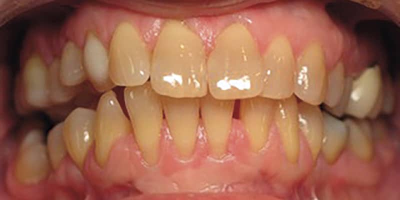
Drs. Barry Glassman, Don Malizia, and Daniele Manfredini discuss various viewpoints on occlusion, pain, and dysfunction
Introduction
The study of dental occlusion and its potential contribution to pain and dysfunction has been a fascinating topic for generations of dentists who spent careers assuming a rather direct relationship. In 1957, Campbell wrote that while dentists should be proud of their advances in restorative therapy, “their concentration on the restorative aspects of their profession has, to some extent, blinded them to the wide implications of pain.”1 Within this historical framework, clinicians must bear in mind that the tendency in dentistry is to think of occlusion as a static relationship that describes how maxillary and mandibular teeth fit together when the elevators contract and maintain contraction — in other words, maximum intercuspation. This seemingly simple concept of occlusion has evoked significant controversy. Based on the belief or negation that less than ideal occlusion may be responsible for dysfunction of the stomagnathic system, clashes of cultures characterized the second half of the 20th century.
The discussion of the role of mal-occlusion as a cause of temporomandibular disorders (TMDs) has an extensive history in dentistry. An examination of the history of the “evidence” of occlusal concepts reveals how several unproven assumptions about occlusion came into existence and why those concepts continue to be taught as “science.” Exposing these potential myths helps us more accurately appreciate how the goal of an “ideal” occlusion in orthodontic care may in fact be out of perspective.
Empirical history
As far back as 1967, Block wrote: “When a patient has myofascial pains in the area near the temporomandibular joint or pains within the joint, the dentist should suspect the presence of occlusal disharmonies.”2 Willie B. May, while researching over 120 chronically ill patients with his physician colleague, Lees, concluded the following about occlusal therapy: “When this treatment is fully researched and understood, it will be capable of revising every diagnosis, treatment procedure, and prognosis in the medical world.”3 In 1988, Fonder wrote of a “Dental Distress Syndrome” describing an etiology of malocclusion. According to Fonder, the resulting pathological structural response to that malocclusion is responsible not only for TMD, but also for neurologic pain, otalgia, visceral symptoms, gynecological symptoms, and various “general symptoms.”4
The orthodontic concept of ideal occlusion being a contributing factor to general health dates back to the mid-1800s with a legendary dentist from Philadelphia, Dr. William Gibson Arlington Bonwell, who was influenced by Freemasonry and religion. Ackerman, et al., report through Bonwell, and later through his student, Angle, that the “science of occlusion emerged from a pseudoscientific tradition already characterized in the 19th century as composed merely of so-called facts connected together by misapprehension under the disguise of principles, and that from the beginning, there were strong overtones of religious belief in the concept of occlusion.”5
The work of Bonwell and Angle led to the belief that ideal orthodontic treatment was the basis of normal dental function and oral health. That myth, despite overwhelming evidence to the contrary over the past 20 years, continues to haunt the dental profession. The deflection to the assumption of purported influence on body posture is just the most recent example. Reid and Greene have outlined the role of conservative therapy in the diagnosis and treatment of orofacial pain and joint dysfunction.6 The likelihood of these well-advised conservative therapeutic principles being followed and understood by dentists other than orofacial pain experts will remain limited until these myths are appropriately dispelled.
In 1973, Roth, an orthodontist, references a series of prosthetic journals and low-quality studies and concludes that it is “generally accepted that the occlusion should ideally be in harmony with the closure and movement patterns that are determined and dictated by the temporomandibular articulations.” His study of only seven post-orthodontic patients demonstrated the presence of TMD symptoms that were eliminated by occlusal treatment. The “control group” of two patients had no postoperative symptoms and no reported occlusal interferences or balancing contacts. This led to the conclusion that “occlusion may play a more important role in the production of the temporomandibular pain-dysfunction syndrome than heretofore ascribed to it.”7
Occlusion continues to be emphasized in a 1973 paper in which Dawson reports, without references, “In my clinical experience, tic douloureux is almost always a misdiagnosis. It is usually nothing more than a classical TMJ syndrome and can be resolved by occlusal therapy.” He further writes, “The pain of TMJ syndrome is almost always resolvable in a matter of minutes once the occlusion has been refined as well as possible.”8 This unfortunate approach has led many dentists to assume that when there is a non-odontogenic facial pain pattern, the resolution will be in the “perfection” of the occlusion, and when an occlusal adjustment does not result in pain relief, the follow-up assumption will be that the occlusal adjustment was not done perfectly. This engineer-like approach to oral rehabilitation still permeates dentistry through the so-called study of gnathology and its derivates — a branch that is not even considered a medical subject heading for Medline search. Failure to consider the implications of nervous system physiopathology and the role of psychological features in the genesis of “pain and dysfunction” are just two of the reasons why gnathological approaches may backfire.

Dental school education of TMD, occlusion, and bruxism
This long history of poorly done research leading to an acceptance of a direct causal relationship between occlusion and TMD without caveat has led to the entrenchment of the concept that “bad bites cause pain.” While it appears that there is limited teaching of occlusion in predoctoral programs, there is no question that students graduate with an awareness that occlusion is important, and that malocclusion can be a cause of TMD. The basic occlusal principles of balanced and bilaterally even contact, canine rise, and anterior guidance are taught at the predoctoral level, making every dentist aware of how important these principles are when performing restorative dentistry. Dentists are trained to avoid creating an occlusal contact that is not in harmony with the existing occlusion. Examples of occlusal prematurities leading to acute pain have further strengthened the concept of causation through confirmation bias.
Despite the emphasis on the principles of occlusion, the basic dental education has not included validated mechanisms of the assumed “causal role” of occlusion in TMD. Predoctoral students report an even more limited curriculum of evidence-based principles on the diagnosis and treatment of TMD and orofacial pain that move past that formerly believed primary causal role of occlusion.9 Young graduates with a limited knowledge of both occlusion and TMD are soon faced with the need to learn more about TMD and facial pain in their new role as dentists. Unfortunately, their limited education in “occlusion and TMD” leaves them vulnerable to those still teaching a direct causal relationship between occlusion, joint dysfunction, and orofacial pain. Logic in the absence of science often prevails.
Competing concepts of occlusion have been at the center of the conflicting TMD “camps” over the years. The difference in “camps” is often related to “joint position,” but they all seem to erroneously accept the role of occlusion as critical and a causal contributing factor in the development of TMD.
Ignored evidence
The controversy continues despite the recent evidence showing limited if any contribution of the static occlusal relationships to pain and dysfunction. The historical acceptance of the causal role of occlusion has been so generally accepted that the mechanism is not fully addressed or questioned by students and dental professionals alike. At the heart of the problem is that many “facts” about occlusion have been developed empirically and passed down as truths.10-12 These “truths” are so cemented into the knowledge base of dentistry that there is great resistance, in spite of the current evidence, to reconsider their validity.
Any theory that involves how occlusion affects the cranio-mandibular-cervical system includes the role of bruxism. There is the tendency to envision the teeth and arches as articulated upper and lower models always fitting together, i.e., occluding. In reality, the occlusal-dental inter-arch contact only occurs about 20 minutes in a 24-hour period.12 In addition to that, there is no evidence that such time is even spent with full contacts between the arches. Bruxing events with tooth contacts can then become the time when occlusion could have the greatest effect on the cranio-mandibular-cervical system.
The future of orthodontic care as an essential specialty of dentistry is not dampened by the realization that the goal of orthodontic care is not occlusally related.
The theory that bruxism is caused by “occlusal interferences” dates back to Karolyi.13 It became an essential part of dental education with Ramfjord’s conclusions in 1961 that occlusal interferences were causal for pain because “Clinically all patients experienced relief of pain and discomfort after complete occlusal adjustment.”14,15 These were poorly designed studies by today’s standards without a control group and are examples of inductive reasoning that has been used to generalize findings far beyond their validity.
Despite the preponderance of the evidence that bruxism is centrally mediated and not the result of any specific occlusal scheme, the causal myth continues.16 This long history suggests that bruxism is the result of malocclusion, and therefore, many patients have been referred for orthodontics or occlusal equilibration to correct their occlusion in order to control their bruxism. This continues to be taught despite the studies that have shown relief from mock equilibration without removing “interferences.”17
The concept of the role of an ideal occlusion and the “fact” that ideal occlusion is critically important to a pain-free and normally functioning trigeminal system remained an assumption in dentistry without any level of evidence beyond empirical observation. In the early 1970s, the National Institute of Dental Research and the National Council of the National Academy of Sciences assigned three independent panels of orthodontic experts to evaluate research related to malocclusion. They essentially concluded that without a good definition of ideal or accepted variations of ideal occlusion, no direct correlations can be made between occlusion and dysfunction.18
In 1976, Weinberg questioned both the concept of “centric relation” and occlusion in the development of joint dysfunction, suggesting that treatment response tended to vary from patient to patient, and that the occlusal scheme is dependent more on the preferences of the dentist, rather than what could be considered required for that unique patient.19 From 1982, the evidence separating occlusal schemes from causation of TMD begins to prevail. In 1982, Graham and Buxbaum conclude that “treatment modalities considered within the first 6 weeks should be conservative and reversible to eliminate or decrease myofascial trigger zones and their area of referred pain. Alteration of the existing occlusion and maxillomandibular relations may be adjusted with caution if necessary.”19 Bush goes a step further in 1984, reporting that “occlusal adjustment appears unsatisfactory as a modality for management of pain.”20
By as early as 2001, Greene notes that the etiological theories on temporomandibular disorders have progressively shifted from peripheral to central factors.16 Despite the fact that some investigators continue to suggest that there is a higher prevalence of TMD in patients with malocclusion as opposed to the “normal population,” “the literature on the effects of orthodontic treatment supports the neutral effects on the temporomandibular joint.”21 The literature support for the lack of a direct relationship between TMD and malocclusion is, in fact, overwhelming. Okeson initially writes that both psychological stress and occlusal interferences were causes of bruxism, thus connecting occlusion as a direct cause of TMD.22 In 2015, Okeson discusses the evolution of the science and reports that “the data did not suggest that orthodontic therapy was a significant risk factor for the development of TMD.”23
Michelotti’s excellent study in 2005 demonstrated that occlusal interferences did not create any pain symptoms in healthy females.24 Manfredini reports that a systematic review (Dworkin) “concluded that there are insufficient research data on the relationship between active orthodontic intervention and TMD on which to base our clinical practice.”22 Another systematic review by McNamara reviews much of the quality evidence and confirms that malocclusion is not a cause of TMD.26 Manfredini, et al., in a retrospective study on a huge sample of over 600 patients concludes that “… our findings support the view that orthodontics is TMD-neutral.”22 While some investigations continue to suggest the importance of various occlusal schemes and joint positions in the development of TMD symptoms,7,27,28 the vast majority of high-level evidence supports the disconnect between occlusion and the development of TMD signs and symptoms. As Greene states,” A clear implication from all those studies is that orthodontic therapy is not required to resolve most TMD cases, regardless of the various occlusal imperfections that may exist in each patient.”29
Summary
The belief that malocclusion is the most likely direct “cause” of TMD has a long history in dentistry. Alvin Toffler has written, “The illiterate of the 21st century will not be those who cannot read and write, but those who cannot learn, unlearn, and relearn.”30 Unlearning is difficult. Restorative gurus continue to teach the direct relationship between malocclusion and pain and dysfunction. Confirmation bias continues to support the long history of empirical evidence. Occlusal adjustments and orthodontics continue to be recommended as first line therapy for patients with various forms of TMD.
There is no better way to summarize the relationship of malocclusion and TMD than the recent findings of the National Academies of Sciences.
“The early treatment of malocclusion through orthodontic treatments was previously considered a viable preventive treatment for TMD. However, the evidence was clear decades ago that orthodontic repositioning of teeth does not prevent the onset of TMD.31 Nevertheless, some dentists have the outdated belief that orthodontic treatment will prevent TMD … [and] … Although commonly suggested as a potential cause, no studies have implicated orthodontic treatment in the development of a TMD.”18
Despite the preponderance of the evidence that clearly separates the state of the occlusion from a direct cause of TMD, patients continue to be referred to orthodontists for orthodontic care to either correct or prevent TMD. Patients are being advised to proceed with dental restorative and prosthetic care to treat or prevent pain or dysfunction. “It is disappointing that the notion that occlusion causes temporomandibular pain [TMD, persistent orofacial pain]persists.”32
In 2005, Rinchuse, et al., commented concerning the continued teaching of an occlusal causation of TMD and thus the support of orthodontic treatment for pain resolution. They wrote: “How can the scientific evidence-based occlusion/TMD knowledge point so clearly in one direction, but dentists and orthodontists ignore this information and practice in a totally different direction.”33
The role of orthodontics today and beyond
Despite the preponderance of the evidence clearly separating “occlusion” from either pain or temporomandibular joint dysfunction, the role of orthodontic therapy has become even more essential. Rather than having an emphasis on how the teeth occlude when the elevator muscles contract and maintain contraction in a nonfunctional posture, a posture that results in dental contact approximately 20 minutes in a full 24-hour period,34 orthodontics of today has the loftier goal of improving craniofacial esthetics with improved function. The orthodontist of tomorrow won’t have glass cases filled with models in ideal occlusion, but walls filled with photos of faces with improved profiles and smiles.
Orthodontic therapy guiding early growth and development can play a major role in the treatment of sleep-disturbed breathing in the young child. “Craniofacial growth influence by genetic inheritance and functional factors can have an impact on general health.”34 Preliminary studies have suggested that orthodontic treatments, such as maxillary expansion or mandibular advancement with functional appliances, may be effective in handling pediatric snoring and OSA.35 Guillminault, et al., demonstrated the effectiveness of the combination of adenotonsillectomy and maxillary rapid expansion in the resolution of pediatric OSA, while treatment of either modality without the other was less likely to be effective.36
The future of orthodontic care as an essential specialty of dentistry is not dampened by the realization that the goal of orthodontic care is not occlusally related. The future is in fact brightened by the orthodontic potential to improve the quality of life by playing a role in the improvement of both esthetics and function. The massive improvement in esthetics can increase the sense of confidence and self-worth of their patients. The significantly increased health resulting from improved sleep with the correction of sleep disturbed breathing has the potential not only to improve the quality of life but also to add years to one’s life expectancy.37
It is the opinion of the authors that the orthodontic community should embrace these evidenced-based changes. This is a very exciting model change for the entire profession.
Besides TMD and occlusion, Drs. Glassman and Malizia have also written for Orthodontic Practice US on the topic of Sleep Medicine. Read their basic review of the subject here: https://orthopracticeus.com/ce-articles/basic-principle-review-of-sleep-medicine/
References
- Campbell JN. Extension of the temporomandibular joint space by methods derived from general orthopedic procedures. J Pros Dent. 1957;7(3):386-399.
- Block LS. Diagnosis of occlusal discrepancies that cause temporomandibular joint or myofacial pain. J Prosthet Dent. 1967;17(5):488-489.
- Fonder AC. The Dental Distress System Quantified. 1988.
- Ackerman JL, Ackerman MB, Kean MR. A Philadelphia fable: how ideal occlusion became the philosopher’s stone of orthodontics. Angle Orthod. 2007;77(1):192-194.
- Reid KI, Greene CS. Diagnosis and treatment of temporomandibular disorders: an ethical analysis of current practices. J Oral Rehabil. 2013; 40(7):546-561.
- Roth RH.Temporomandibular pain-dysfunction and occlusal relationships. Angle Orthod. 1973;43(2):136-153.
- Dawson PE. Temporomandibular joint pain-dysfunction problems can be solved. J Prosthet Dent. 1973;29(1):100-112.
- Steenks MH. The gap between dental education and clinical treatment in temporomandibular disorders and orofacial pain. J Oral Rehabil. 2007. 34(7):475-477.
- Guichet NF. Biologic laws governing functions of muscles that move the mandible. Part III. Speed of closure–manipulation of the mandible. J Prosthet Dent. 1977;38(2):174-179.
- Guichet NF. Biologic laws governing functions of muscles that move the mandible. Part II. Condylar position. J Prosthet Dent. 1977;38(1):35-41.
- Guichet NF. Biologic laws governing functions of muscles that move the mandible. Part I. Occlusal programming. J Prosthet Dent. 1977;37(6): 648-656.
- Graf H. Tooth Contact Patterns in Mastication. J Prosthet Dent. 1963;13(6):1055-1066.
- Karolyi M. Beobachtungen uber Pyorrhea alveolaris. Oesterr-Ungar Bierteljahrsschrift Zahnheilkunde. 1901.
- Ramfjord, S.P., Dysfunctional temporomandibular joint and muscle pain. J Prosthet Dent. 1961;11(2):353-374.
- Ramfjord SP. Bruxism, a clinical and electromyographic study. J Am Dent Assoc. 1961;62:21-44.
- Greene CS. The etiology of temporomandibular disorders: implications for treatment. J Orofac Pain. 2001;15(2):93-105.
- Goodman P, Greene CS, Laskin DM. J Am Dent Assoc. 1976;92(4):755-758.
- NASEM. Temporomandibular Disorders: Priorities for Research and Care. National Academies of Sciences, Engineering, and Medicine. Washington, DC: The National Academies Press; 2020.
- Weinberg LA. Temporomandibular joint function and its effect on concepts of occlusion. J Prosthet Dent. 1976;35(5):553-566.
- Graham MM, Buxbaum J, Staling LM. A study of occlusal relationships and the incidence of myofacial pain. J Prosthet Dent. 1982;47(5):549-555.
- Bush FM. Occlusal therapy in the management of chronic orofacial pain. Anesth Prog. 1984;31(1):10-16.
- Manfredini D, Stellini E, Gracco A, et al. Orthodontics is temporomandibular disorder-neutral. Angle Orthod. 2016;86(4):649-654.
- Okeson JP. Etiology and treatment of occlusal pathosis and associated facial pain. J Prosthet Dent. 1981;45(2):199-204.
- Okeson JP. Evolution of occlusion and temporomandibular disorder in orthodontics: Past, present, and future. Am J Orthod Dentofacial Orthop. 2015;147(Suppl 5):S216-S223.
- Michelotti A, Farella M, Gallo LM, et al. Effect of occlusal interference on habitual activity of human masseter. J Dent Res. 2005;84(7):644-648.
- McNamara JA Jr, Seligman DA, Okeson JP. Occlusion, Orthodontic treatment, and temporomandibular disorders: a review. J Orofac Pain. 1995;9(1):73-90.
- Alanen, P., Occlusion and temporomandibular disorders (TMD): still unsolved question? J Dent Res. 2002;81(8):518-519.
- Monaco A, Petrucci A, Marzo G, et al. Class II malocclusion on the kinesiographic pattern of young adolescents: a case-control study. 2013;14(2):131-134.
- Greene CS, Galang-Boquiren MTS, Bartilotta BY. Orthodontics and the temporomandibular joint: What orthodontic providers need to know. Quintessence Int. 2017;48:799-808.
- Toffler A. Future Shock. New York:Random House; 1970.
- McNamara JA Jr. Orthodontic treatment and temporomandibular disorders. Oral Surg Oral Med Oral Pathol Oral Radiol Endod. 1997;83(1): 107-117.
- Zakrzewska JM. Education is better than splints in temperomandibular pain. BMJ. 2012;345:e7447.
- Rinchuse DJ, Rinchuse DJ, Kandasamy S. Evidence-based versus experience-based views on occlusion and TMD. Am J Orthod Dentofacial Orthop. 2005;127(2):249-254.
- Graf H. Bruxism. Dent Clin North Am. 1969;13:659-665.
- Huynh NT, Desplats E, Almeida FR. Orthodontics treatments for managing obstructive sleep apnea syndrome in children: A systematic review and meta-analysis. Sleep Med Rev. 2016;25:84-94.
- Guilleminault C, Quo S, Huynh NT, Li K. Orthodontic expansion treatment and adenotonsillectomy in the treatment of obstructive sleep apnea in prepubertal children. Sleep. 2008;31(7):953-957.
- Jordan AS, McSharry DG, Malhotra A. Adult obstructive sleep apnoea. Lancet. 2014;383:736-747.
Stay Relevant With Orthodontic Practice US
Join our email list for CE courses and webinars, articles and mores

 Barry Glassman, DMD, maintained a private practice in Allentown, Pennsylvania, which was limited to orofacial pain and dental sleep medicine. He is a Diplomate of the Board of the American Academy of Craniofacial Pain, a Fellow of the International College of Craniomandibular Orthopedics, and a Diplomate of the American Academy of Pain Management. He is a Diplomate of the Board of the American Academy of Dental Sleep Medicine. He teaches and lectures internationally on orofacial pain, joint dysfunction, and sleep disorders.
 Don Malizia, DDS, limits his practice to orofacial pain and sleep-disturbed breathing at the Allentown Pain and Sleep Center inWilkes-Barre, Pennsylvania. Among his recent publications, coauthored with Dr. Glassman, are “The Effect of Regional Anesthetic Sphenopalatine Ganglion Block on Self-Reported Pain in Patients With Status Migrainosus” in Headache and “The Curious History of Occlusion in Dentistry” in Dentaltown.
 Daniele Manfredini, DDS, PhD, MSc, received his DDS from the University of Pisa, Italy, in 1999, a MSc in Occlusion and Craniomandibular Disorders in 2001 from the same University, a PhD in Dentistry from the ACTA Amsterdam, The Netherlands, in 2011, and a Post-Graduation Specialty in Orthodontics from the University of Ferrara, Italy, in 2017. He achieved the Diplomate Status from the American Academy of Orofacial Pain in 2021. He was a clinical fellow at the Section of Prosthetic Dentistry, Department of Neuroscience, University of Pisa, Italy until 2005. From 2006 to 2016, Daniele Manfredini has been Assistant Professor at the School of Dentistry and coordinator of the research projects at the TMD Clinic, Department of Maxillofacial Surgery, University of Padova, Italy. Since 2017, Daniele Manfredini has held teachings in Oral Physiology and in Clinical Gnathology at the School of Dentistry, University of Siena, Italy. On January 2017, the Italian Ministry of University and Research (MIUR) appointed him as a Full Professor by scientific merit at the age of 41. Daniele Manfredini authored more than 210 papers in the field of bruxism and temporomandibular disorders in journals indexed in the Medline database (Scopus H-index=42). He also edited, among others, the book Current Concepts on Temporomandibular Disorders (Quintessence Publishing, 2010). Since 2018, Daniele Manfredini has been a Member and Coordinator of the Bruxism Consensus Panel within the International Association for Dental Research, which works on the updated definition and classification strategies for bruxism.
