CEU (Continuing Education Unit): 2 Credits
Educational aims and objectives
This self-instructional course for dentists looks at the critical role of the dental professional in the early detection of head and neck cancers.
Expected outcomes
Orthodontic Practice US subscribers can answer the CE questions by taking the quiz online to earn 2 hours of CE from reading this article. Correctly answering the questions will demonstrate the reader can:
- Recognize the importance of the Conventional Visual and Tactile Examination (CVTE) in routine dental visits for early detection of head and neck cancers.
- Identify and describe each component of a systematic extraoral and intraoral head and neck exam.
- Understand the current evidence-based guidelines that support the implementation of CVTE in dental practice.
- Apply practical strategies for incorporating the CVTE into clinical workflows to enhance patient outcomes.

Dr. Brett Gilbert and Jonathan Gegerson provide a call to action for head and neck screening in dental practice
Abstract
This article underscores the critical role of the dental professional in the early detection of head and neck cancers through a systematic Conventional Visual and Tactile Examination (CVTE). Inspired by the personal story of my close friend, Jonathan, whose cancer diagnosis may have been expedited by routine dental screening, this article explores the components of the head and neck exam, its clinical rationale, and the current evidence supporting its implementation as a standard of care in dental settings.
Jonathan’s story in his own words: a missed opportunity at the dental chair
There I sat waiting for the doctor to examine my concern of a lump on the right side of my neck. I assumed it was nothing to be concerned about. The doctor entered the room, and I showed her the area. She felt around my neck on both sides, felt under my arms, and then asked a couple questions. “Have you had any dental work recently?” I responded, “No.” She then asked, “Have you had any infections in the mouth, or a root canal or anything?” Once again, my response was, “No.” She then looked at me and said, “It may be cancer.” I was stunned, and my body and mind froze in that moment, as that was the last thing I thought I was going to hear. Other than the lump on my neck, I was in perfect health. I worked out constantly, ate a Keto diet, never smoked, and limited all fatty foods and alcohol. There was no reason I should have cancer — at least that is what I thought.
Since my initial diagnosis in May 2019, I have received over 200 rounds of chemotherapy, 67 sessions of radiation, and six surgeries. The side effects from all these treatments have been overwhelming at times. I lost my ability to chew and must be on a soft, liquid diet. My mouth opens just wide enough for a spoon. I was on a feeding tube for 6 months. I have constant lymphedema of my face, tongue, and throat which creates speaking problems, swallowing concerns, and at times, vision issues. I have radiation scars on my face, and one of my vocal cords was paralyzed during my recent 17-hour surgery, which was to help correct lymphedema as my airway was being constricted. I ended up in the ICU twice due to airway constriction. My body was literally suffocating itself.
I am the second patient to receive the type of surgery I had for head and neck lymphedema. My recovery took 7 months, and for two of those months, I was in the hospital. I share this because there were opportunities to catch the cancer sooner. Those opportunities were in my own power as well as the power of my dentist and hygienist. I mention my hygienist and dentist as I would see them regularly — at least every 6 months. If the cancer was caught sooner, it may have prevented such an extensive treatment plan, limited all my side effects, and resulted in a better lifestyle after treatment.
I would rather have been told there was a concern 3 or 6 months before by my dentist or hygienist and addressed the concern with my primary doctor immediately. Early detection results in better patient outcomes. I was informed by my oncologist that out of 4 million people, 10 to 15 people get the type of cancer I was diagnosed with: Salivary Duct Carcinoma HER2 Positive. I was also told that I have been beating the odds, and I am one of a kind. I would rather be many of a kind when it comes to living through and after cancer. This is one of my motivations to encourage all hygienists and dentists to perform a cancer screening exam that includes the neck. Most people with my diagnosis are not here to tell their story. I intend to purposefully speak for them and myself as I encourage all dental professionals to perform a head and neck exam on every adult patient, every time they see them!
The clinical imperative
The dental setting provides a unique, often untapped opportunity to detect early signs of head and neck malignancies. The Conventional Visual and Tactile Examination (CVTE) is a simple, low-cost, evidence-based procedure that can uncover abnormalities before they become life-threatening.
The American Dental Association (ADA) recommends that clinicians perform a systematic CVTE for all adult dental patients, including inspection and palpation of the face, neck, and regional lymph nodes to detect tissue changes, masses, or asymmetry.1 This recommendation is echoed by the American Academy of Otolaryngology–Head and Neck Surgery, which emphasizes evaluation of the skin, salivary glands, thyroid, and lymph nodes using bimanual palpation techniques.2
Despite this guidance, implementation in general practice remains inconsistent.3
Step-by-step: performing the Conventional Visual and Tactile Examination (CVTE)
General observation
Observe facial symmetry, skin color/lesions, swelling, and visible masses. Note signs of asymmetry, facial droop, or skin changes.
Lymph node palpation
Preauricular and postauricular, submental and submandibular, cervical chain (anterior and posterior), supraclavicular nodes. Use gentle, circular pressure with the pads of your fingers, bilaterally when appropriate.
Salivary glands
Palpate parotid, submandibular, and sublingual glands for enlargement, tenderness, or firmness (Figure 1).
Thyroid gland
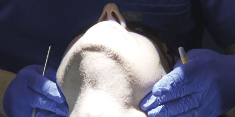
Visually inspect for enlargement while the patient swallows. Palpate the thyroid gently from behind the patient using both hands (Figure 2).
Floor of mouth (Bimanual palpation)
Place one gloved hand inside the mouth and one under the chin. Gently compress to assess for firm masses or nodularity.
Intraoral exam
Thorough inspection of the lips, buccal mucosa, gingiva, hard/soft palate, tongue (dorsal, lateral, and ventral), and oropharynx. Use gauze to pull and manipulate the tongue for complete visualization (Figures 3-4).
A case from my chair: CVTE in action
While Jonathan’s story reflects the devastating consequences of a missed opportunity for early detection, I also carry with me the opposite experience — one in which a thorough Conventional Visual and Tactile Examination (CVTE) led to the early diagnosis of a life-threatening malignancy.
In 2016, a patient presented to my endodontic clinic for evaluation of a sore area on the palatal tissue adjacent to tooth No. 14 (Figure 5). As with every patient encounter, I performed a standard diagnostic endodontic evaluation alongside a systematic CVTE.
Clinical Findings
- Percussion: Negative
- Palpation: Positive on the palatal surface of tooth No. 14
- Periodontal Probing: Within normal limits
- Mobility: Within normal limits
- Pulpal Sensitivity (Cold): Normal response
These results pointed toward an endodontic diagnosis for tooth No. 14 of normal pulp with symptomatic apical periodontitis (due to a positive finding of palpation on the palate). But what caught my attention was not the tooth — it was the tissue.
During the CVTE, I noted a small raised swelling on the palatal surface (Figure 6) and a separate white nodular lesion on the buccal gingiva adjacent to tooth No. 14 (Figure 7). Importantly, these findings did not align with any odontogenic pathology typically associated with tooth No. 14. That clinical inconsistency provided the moment to pause and widen the diagnostic lens.
Referral and diagnosis
Given the suspicious nature of these findings, I referred the patient to an oral and maxillofacial surgeon for biopsy. Oral and Maxillofacial (OMS) surgeons are often the first line of defense for dentists who may be unsure about a diagnosis that does not appear to be odontogenic in origin. The term “non-leo” refers to a lesion of non-endodontic origin. As a dental specialist, determination that findings are not adding up to a diagnosis of dental origin must be referred immediately.
OMS is the first line of referral for dentists as these specialists often have had extensive medical and hospital training. A dentist must understand that when they detect an irregular finding, no matter how small, it is critical to make this referral. Immediate referral will shorten the time between detection and diagnosis.
In many cases, the referral may seem like it was excessive if the OMS does not diagnose a problem. As clinicians, we should never allow doubt to creep into our minds by thinking that the finding is likely not significant. In fact, a non-significant diagnosis is the hope of the referral! The conversation we have with a patient in this moment should be calm in tone and decisive. I often will have my clinical team call the OMS office to set up a consultation appointment while the patient is still in our office. We must consider that a patient may listen to your concern but not act by making an appointment which could decrease the chances of a good outcome if a serious medical diagnosis is confirmed.
This patient was compliant and presented to the OMS the next day. The OMS performed their own examination and determined that a biopsy was necessary. The biopsy results revealed:
“Diffuse large B-cell lymphoma of the left maxillary sinus, germinal center phenotype” (Figure 8).
The patient was subsequently referred to oncology and underwent successful treatment for which they were extremely grateful that the detection of such a devastating systemic illness was detected early in the dental chair.
Clinical takeaway
This case illustrates the critical importance of integrating CVTE with routine diagnostic protocols. If I had focused solely on the dental findings, the underlying malignancy may have been missed. It was the intentional soft tissue assessment through CVTE that revealed the warning signs.
When something doesn’t “add up” between tooth-level findings and soft tissue presentation, it’s time to:
- Pause and reassess
- Expand the differential beyond odontogenic causes
- Refer promptly to an oral and maxillofacial surgeon
Head and neck cancers often masquerade as benign or dental conditions — or remain entirely silent. A few extra moments of systematic examination can create a critical bridge to life-saving intervention.
Evidence basis: why it matters
CVTE remains the gold standard in dental practice for early detection of head and neck malignancy,1,2,3 according to multiple high-level guidelines and reviews. Some literature shows that adjunctive screening tools (e.g., fluorescence imaging, salivary biomarkers) have not demonstrated sufficient evidence to replace or supplement CVTE in routine practice.4,5 However, any device or screening tool that reminds a clinician how important it is to conduct these exams are worthy. CVTE should be considered a critical part of the dental examination and can be used in conjunction with any other screening tool that a clinician feels is valuable.
Recent systematic reviews confirm that visual inspection and palpation of the head, neck, and lymph nodes are the most frequently implemented and most reliable methods for early cancer detection in dental settings.6,7
Emerging test for high detection of oro-pharyngeal cancers is under investigation
Recent studies, including Das, et al., (2024, 2025),8,9 demonstrate that the HPV-DeepSeek assay — using whole-genome sequencing of circulating tumor HPV DNA (ctHPV-DNA) — achieves high sensitivity (up to 96–99%) and specificity (up to 99%) for detecting HPV-positive oropharyngeal squamous cell carcinoma, with detection possible up to 7.8–10 years before clinical diagnosis in some cases.8,9,10,11 This supports the claim that HPV-DeepSeek is a highly accurate, non-invasive liquid biopsy with potential for early cancer detection.
However, the clinical utility of HPV-DeepSeek as a routine screening tool in the dental setting remains investigational11,12,13 While the test shows promise for early detection and could theoretically complement head and neck examinations in dentistry, there is currently no guideline or regulatory approval for its use in routine screening or as a standard adjunct in dental practice. The National Comprehensive Cancer Network (NCCN) guidelines emphasize that blood-based ctHPV-DNA assays are not yet part of standard screening or diagnostic protocols and that their performance and actionable implications outside of clinical trials are still being evaluated.11
Closing thoughts: a moral and clinical duty
For Jonathan, the absence of a head and neck exam may have cost him an earlier diagnosis, a simpler treatment, and a better quality of life. He has beaten the odds and desperately wants to share his story as a motivation and wake up call for dental professionals. His story is a call to action — for dentists and hygienists to go beyond the minimum, to reclaim our critical role in early cancer detection, and to never underestimate the power of a few minutes of intentional, hands-on examination.
Working in conjunction with other dental specialists to get a second opinion or to perform more advanced testing procedures, such as a biopsy or other imaging, represents best practice in dentistry. As dental professionals, we often have more opportunities to see and examine our patients than a primary physician. We cannot become complacent and avoid performing the CVTE just because the patient appeared healthy at prior visits. Thorough review of patient health history, medications, and any acknowledgement of oral habits such as smoking, oral placement of tobacco, and even consumption of alcohol should further motivate clinicians to remember to perform this life-saving examination.
As dentists, we do not have to take on the burden of making a definitive diagnosis on cases that present with abnormal findings. Our duty and responsibility are only to make a timely and appropriate referral for further examination and testing. By making the CVTE a routine, non-negotiable part of every adult dental visit, we honor our patients, our profession, and the principle that dentistry is, at its core, a healing art.
Besides CVTE, some clinicians are adding technology to their tools for detecting oral cancer. Read some research on Velscope tissue fluorescence technology here: https://orthopracticeus.com/industry-news/successful-results-latest-clinical-research-using-velscope-tissue-fluorescence-technology/.
References
REFERENCES:
- Lingen MW, Abt E, Agrawal N, Chaturvedi AK, Cohen E, D’Souza G, Gurenlian J, Kalmar JR, Kerr AR, Lambert PM, Patton LL, Sollecito TP, Truelove E, Tampi MP, Urquhart O, Banfield L, Carrasco-Labra A. Evidence-based clinical practice guideline for the evaluation of potentially malignant disorders in the oral cavity: A report of the American Dental Association. J Am Dent Assoc. 2017 Oct;148(10):712-727.e10. doi: 10.1016/j.adaj.2017.07.032.
- Pynnonen MA, Gillespie MB, Roman B, Rosenfeld RM, Tunkel DE, Bontempo L, Brook I, Chick DA, Colandrea M, Finestone SA, Fowler JC, Griffith CC, Henson Z, Levine C, Mehta V, Salama A, Scharpf J, Shatzkes DR, Stern WB, Youngerman JS, Corrigan MD. Clinical Practice Guideline: Evaluation of the Neck Mass in Adults Executive Summary. Otolaryngol Head Neck Surg. 2017 Sep;157(3):355-371. doi: 10.1177/0194599817723609.
- Pynnonen MA, Gillespie MB, Roman B, Rosenfeld RM, Tunkel DE, Bontempo L, Brook I, Chick DA, Colandrea M, Finestone SA, Fowler JC, Griffith CC, Henson Z, Levine C, Mehta V, Salama A, Scharpf J, Shatzkes DR, Stern WB, Youngerman JS, Corrigan MD. Clinical Practice Guideline: Evaluation of the Neck Mass in Adults. Otolaryngol Head Neck Surg. 2017 Sep;157(2_suppl):S1-S30. doi: 10.1177/0194599817722550.
- Moyer VA; U.S. Preventive Services Task Force. Screening for oral cancer: U.S. Preventive Services Task Force recommendation statement. Ann Intern Med. 2014 Jan 7;160(1):55-60. doi: 10.7326/M13-2568.
- Huber MA. Adjunctive Diagnostic Techniques for Oral and Oropharyngeal Cancer Discovery. Dent Clin North Am. 2018 Jan;62(1):59-75. doi: 10.1016/j.cden.2017.08.004. Epub 2017 Oct 16.
- Louredo BVR, de Lima-Souza RA, Pérez-de-Oliveira ME, Warnakulasuriya S, Kerr AR, Kowalski LP, Hunter KD, Prado-Ribeiro AC, Vargas PA, Santos-Silva ARD. Reported physical examination methods for screening of oral cancer and oral potentially malignant disorders: a systematic review. Oral Surg Oral Med Oral Pathol Oral Radiol. 2024 Feb;137(2):136-152. doi: 10.1016/j.oooo.2023.10.005. Epub 2023 Oct 16.
- Sykes EA, Weisbrod N, Rival E, Haque A, Fu R, Eskander A. Methods, Detection Rates, and Survival Outcomes of Screening for Head and Neck Cancers: A Systematic Review. JAMA Otolaryngol Head Neck Surg. 2023 Nov 1;149(11):1047-1056. doi: 10.1001/jamaoto.2023.3010.
- Das D, Hirayama S, Aye L, Bryan ME, Naegele S, Zhao B, Efthymiou V, Mendel J, Fisch AS, Kröller L, Michels BE, Waterboer T, Richmon JD, Adalsteinsson V, Lawrence MS, Crowson MG, Iafrate AJ, Faden DL. Blood-based screening for HPV-associated cancers. medRxiv [Preprint]. 2024 Feb 2:2024.01.04.24300841. doi: 10.1101/2024.01.04.24300841.
- Bryan ME, Aye L, Das D, Hirayama S, Al-Inaya Y, Mendel J, Naegele S, Efthymiou V, Alzumaili B, Faquin WC, Sadow PM, Lin D, Varvares MA, Feng AL, Deschler DG, Chan AW, Paly J, Park JC, Roberts T, Merkin R, Mishra SK, Kröller L, Michels B, Iafrate AJ, Wirth LJ, Adalsteinsson VA, Crowson M, Waterboer T, Mirabello L, Lawrence MS, Guan Z, Fisch AS, Richmon JD, Faden DL. Direct Comparison of Alternative Blood-Based Approaches for Early Detection and Diagnosis of HPV-Associated Head and Neck Cancers. Clin Cancer Res. 2025 Aug 14;31(16):3483-3493. doi: 10.1158/1078-0432.CCR-24-2525.
- National Comprehensive Cancer Network. Head and Neck Cancers. Practice Guideline. Updated August 12, 2025. https://www.nccn.org/guidelines/guidelines-detail? category=1&id=1437.
- Poljak M, Cuschieri K, Alemany L, Vorsters A. Testing for Human Papillomaviruses in Urine, Blood, and Oral Specimens: an Update for the Laboratory. J Clin Microbiol. 2023 Aug 23;61(8):e0140322. doi: 10.1128/jcm.01403-22. Epub 2023 Jul 13.
- Araujo M, Bouassaly J, Farshadi F, Hier M, Mascarella M, Mlynarek A, Alaoui-Jamali M, da Silva SD. Current status of circulating tumor DNA and circulating cell alterations in HPV-associated head and neck cancer. Oral Oncol. 2025 Aug;167:107417. doi: 10.1016/j.oraloncology.2025.107417. Epub 2025 Jun 13.
Stay Relevant With Orthodontic Practice US
Join our email list for CE courses and webinars, articles and mores
