Drs. German O. Ramirez-Yañez and Carlos M. Mejia-Gomez discuss intercepting developing malocclusions as early as possible to reduce the risk of more complicated treatments
Abstract
A clinical case is presented showing a non-syndromic child born with a severely retruded mandible and deep bite. An early intervention permitted improvement of the sagittal and vertical relationships between both maxillaries before the patient enters into the mixed dentition.
[userloggedin]
This clinical case supports the idea that developing malocclusions in children should be intercepted as early as possible in order to reduce the risk of more complicated treatments in the future, as well as preventing other problems that can associate with a deviated or diminished craniofacial growth and development.
 Introduction
At birth, the mandible is positioned distal to the maxilla in a sagittal relation.1 The mandible presents a high growing rate over the first year of life,2 improving the sagittal, transverse, and vertical relationships between both maxillaries.3,4 Some children are born with a severely retruded mandible, which makes their face appear as if they had Pierre Robin syndrome but without the cleft palate and glossoptosis characteristic of that congenital malformation.
The higher potential for mandibular and maxillary growth has been reported to happen over the first 5 years of life.2,5 Even more, a distocclusion at the primary dentition is going to perpetuate or even worsen through the mixed dentition.6,7 Also, a retruded mandible is associated with a retrusive tongue position.8,9 A child with those conditions is at higher risk of developing sleep-related breathing dis-
orders,10 and therefore, an early intervention in a child with a retruded mandible may be beneficial, as it may reduce the severity of the problem and its detrimental effects on the oral functions.6,11
This paper reports a clinical case of a non-syndromic child born with a severely retruded mandible and deep bite. An early intervention permitted improvement of the sagittal and vertical relationships between both maxillaries before the patient enters into the mixed dentition.
Case report
The patient was initially seen at 10 months old since the mother was concerned that he had a small mandible with no chin and was sleeping with the mouth open and breathing noisily. The medical history was not relevant, and he was naturally delivered with no complications. The patient was receiving respiratory therapy. Breastfeeding happened over the first 4 months, and then the mother gave up as she did not produce enough milk. She had been feeding him with formula since then.
The first clinical exam revealed a distal position of the mandible (8 mm), associated with deep bite (OB 100%). The patient had hyperactivity of the mentalis muscles at swallowing and lips unsealed at rest. The initial position of the mandible and the dental occlusion are shown in Figures 1A, 2A, and 3A. At this age, myofunctional therapy was initiated in order to stimulate lip seal. The mother was advised to exercise this area by maintaining the lips together with her fingers for 5 minutes for 3 to 5 times per day. That exercise was practiced over a 3-month period. At 14 months old, the patient was maintaining a lip seal most of the time (Figure 1B).
After maintaining lip seal, the mother was instructed to add other exercises, such as massage on the tongue to stimulate an anterior movement of the tongue and massage on the incisive papilla and mandibular traction with her fingers, bringing the mandible forward. These exercises were also recommended 3 to 5 times per day. Besides that, the mother was instructed to slowly harden the diet by progressively introducing food with fiber, such as carrots, crackers, and meats, into his diet.
At 36 months old, the patient showed some improvement in the sagittal relationship between the maxillaries and the profile (Figure 1C). Another exercise was introduced at this stage. The mother was instructed to place a piece of paper on the side of his mouth and ask the child to bite toward that side.
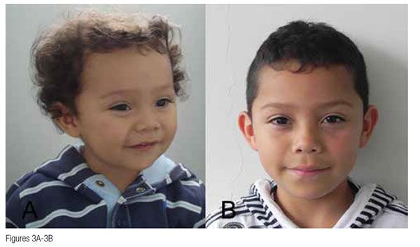
The Planas Direct Tracks (PDTs) were built up when the patient was 42 months old. They were built up on the first primary molars as described by Ramirez-Yañez for disto-occlusion.12 The patient was followed up over the next 12 months, adjusting the PDTs in order to stimulate a forward displacement of the mandible. The overjet and overbite were within normal limits at 48 months old (Figure 2B). Since then, the patient has been wearing a functional removable appliance, the Indirect Planas Tracks, as a retainer waiting for the eruption of the first permanent molars. The patient was instructed to wear the latest appliance 24 hours per day, removing it only for eating. At 5 years old, the patient is showing a normal sagittal, transverse, and vertical relationship for his age (Figure 2B). Also, his profile has improved with the treatment (Figures 1A-1D and Figures 3A-3B).
 Discussion
A disto-occlusion diagnosed in the primary dentition does not improve with natural growth. A longitudinal study reported that conversely, the developmental problem is going to be present in the mixed dentition or even become worse.6 Furthermore, that developmental problem can associate with sleep-breathing disorders,10 which may further affect the growth and development of the child.13
The case presented here was intercepted at an early age in the primary dentition, so growth and development of both maxillaries were stimulated during the period of his life when they expressed the highest growth potential.2,5 In that context, the developmental problem was successfully intercepted, and the mouth was brought to a situation where growth and development can continue within normal limits.11,14
The patient was initially treated with myofunctional exercises and diet guidance, which help to improve the relationship between the maxillaries. Also, that treatment improved the activity of the masticatory and facial muscles, making them able to hold the mandible in a centric position.15 The improvement in the maxillaries’ relationship was associated with an improvement in the oral functions, such as swallowing, lip seal, and tongue posture at rest16,17, which can make the results of the treatment more stable.18 Later, when the patient was more cooperative, a simple, fixed technique was designed for primary dentition. The PDTs were introduced to further stimulate the relationship of the maxillaries in a sagittal and vertical way, with the intent of guiding the craniofacial growth and development.
In this case, early treatment improved the relationship between both maxillaries and guided the craniofacial growth and development. Thus, all the tissues composing the oral system can continue expressing their highest growing potential over the following stages. In other words, the message received by the trigeminal nerve through the mechanoreceptors in the periodontal ligament is sent to the brain, which in turn will change the activity of the mandibular and facial muscles. This produces better loading of the craniofacial bones, including both maxillaries.14,19,20
In conclusion, the clinical case presented here supports the idea that developing malocclusions in children should be intercepted as early as possible in order to reduce the risk of more complicated treatments in the future, as well as preventing other problems that can associate with deviated or diminished craniofacial growth and development, such as sleep-breathing disorders.
[/userloggedin]
[userloggedout][/userloggedout]
- Smartt JM Jr., Low DW, Bartlett SP. The pediatric mandible: I. A primer on growth and development. Plast Reconstr Surg. 2005;116(1):14e-23e.
- Liu YP, Behrents RG, Buschang PH. Mandibular growth, remodeling, and maturation during infancy and early childhood. Angle Orthod. 2010;80 (1):97-105.
- Kobayashi HM, Scavone H Jr., Ferreira RI, Garib DG. Relationship between breastfeeding duration and prevalence of posterior crossbite in the deciduous dentition. Am J Orthod Dentofacial Orthop. 2010;137(1):54-58.
- Westover KM, DiLoreto MK, Shearer TR. The relationship of breastfeeding to oral development and dental concerns. ASDC J Dent Child. 1989;56(2):140-143.
- Laowansiri U, Behrents RG, Araujo E, Oliver DR, Buschang PH. Maxillary growth and maturation during infancy and early childhood. Angle Orthod. 2013;83(4):563-571.
- Baccetti T, Franchi L, McNamara JA Jr., Tollaro I. Early dentofacial features of Class II malocclusion: a longitudinal study from the deciduous through the mixed dentition. Am J Orthod Dentofacial Orthop. 1997;111(5):502-509.
- Ovsenik M, Farcnik FM, Korpar M, Verdenik I. Follow-up study of functional and morphological malocclusion trait changes from 3 to 12 years of age. Eur J Orthod. 2007;29(5):523-529.
- Bacon WH, Turlot JC, Krieger J, Stierle JL. Cephalometric evaluation of pharyngeal obstructive factors in patients with sleep apneas syndrome. Angle Orthod. 1990;60(2):115-122.
- Yılmaz F, Sağdıç D, Karaçay S, Akin E, Bulakbası N. Tongue movements in patients with skeletal Class II malocclusion evaluated with real-time balanced turbo field echo cine magnetic resonance imaging. Am J Orthod Dentofacial Orthop. 2011;139(5):e415-e425.
- Guilleminault C, Akhtar F. Pediatric sleep-disordered breathing: New evidence on its development. Sleep Med Rev. 2015;24:46-56.
- Ovsenik M. Incorrect orofacial functions until 5 years of age and their association with posterior crossbite. Am J Orthod Dentofacial Orthop. 2009;136:375-381.
- Ramírez-Yañez G. Early treatment of malocclusions: prevention and interception in primary dentition. 2009; 2nd ed:www.kidsmalocclusions.com. Accessed May 27, 2016.
- Huang YS, Guilleminault C. Pediatric obstructive sleep apnea and the critical role of oral-facial growth: evidences. Front Neurol. 2013;3:184.
- Sohn BW, Miyawaki S, Noguchi H, Takada K. Changes in jaw movement and jaw closing muscle activity after orthodontic correction of incisor crossbite. Am J Orthod Dentofacial Orthop 1997;112(4):403-409.
- Maffei C, Garcia P, de Biase N, et al. Orthodontic intervention combined with myofunctional therapy increases electromyographic activity of masticatory muscles in patients with skeletal unilateral posterior crossbite. Acta Odontol Scand. 2014;72(4):298-303.
- Korbmacher HM, Schwan M, Berndsen S, Bull J, Kahl-Nieke B. Evaluation of a new concept of myofunctional therapy in children. Int J Orofacial Myology. 2004;30:39-52.
- Schievano D, Rontani R, Bérzin F. Influence of myofunctional therapy on the perioral muscles. Clinical and electromyographic evaluations. J Oral Rehabil. 1999;26(7):564-569.
- Smithpeter J, Covell D Jr. Relapse of anterior open bites treated with orthodontic appliances with and without orofacial myofunctional therapy. Am J Orthod Dentofacial Orthop. 2010;137(5):605-614.
- Forwood MR. Mechanical effects on the skeleton:are there clinical implications? Osteoporos Int. 2001;12(1):77-83.
- Frost HM. A 2003 update of bone physiology and Wolff’s law for clinicians. Angle Orthod. 2004;74(1):3-15.
Stay Relevant With Orthodontic Practice US
Join our email list for CE courses and webinars, articles and mores
