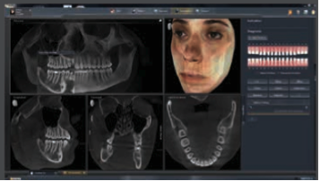
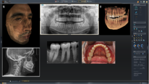
Intuitive software and new features such as patient timeline and lightbox for gathering and displaying images sets a new standard in clinical diagnosis and patient care.

typical clinical procedures.
Already feedback from numerous clinicians nationwide suggests that, with the introduction of SIDEXIS 4 software, Sirona Dental has once again set a new industry standard, this time in clinical diagnosis and patient communication.
Private practitioners Neal Patel, DDS, of Powell, Ohio, and Tarun Agarwal, DDS, of Raleigh, NC, recently discussed how this innovative software, with a sleek modern look, has improved their ability to diagnose and treatment plan and—perhaps most importantly—effectively communicate their findings to patients, who, are then more likely to accept recommended treatment.
Like many practitioners, Dr. Agarwal is concerned about potential downtime associated with any change to an existing system. However, he said, “SIDEXIS 4 takes ease of use to another level. What was previously hidden in the menu bar is now clearly visible in workflow icons arranged to match typical clinical procedures. Our team was using it literally 30 minutes after we installed it.“

Dr. Patel considered the software integration to be seamless and the workflow streamlined, offering the ability to move quickly from the mode of diagnosing an image and transition to patient education, treatment planning and, ultimately, case acceptance. He calls SIDEXIS 4 “the ultimate software of importing and exporting images in various formats,” readily accepting images obtained by intraoral, panoramic, and cephalometric x-rays, 2D and 3D CBCT systems, intraoral cameras, FaceScanner, and more.
Dr. Patel finds especially valuable the visual patient timeline, which enables the clinician or team member to scroll through a complete patient history to select and retrieve desired images based on acquisition date. “Before, if I wanted to view an image taken 2 or 3 years earlier of the current area of concern, I would have to close one image to open another. Now I can just scroll through the timeline for the earlier image seamlessly without having to open up folders and search for specific locations.”

over time offers objective anatomical comparisons.
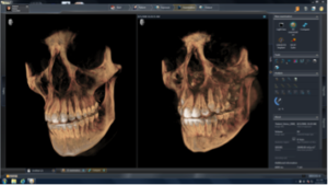
Complementing the timeline, says Dr. Agarwal is the lightbox feature, that, with just a few clicks, enables side-by-side display of multiple images taken over time, offering objective anatomical comparisons of 2D, 3D, intraoral, or camera images. “If we have a scan or image from a few years back, we are able to take that image and compare it to an image we just took.”
Dr. Patel adds that ‘comparing apples to apples’—especially cone beams—to be especially powerful for both diagnosis and patient communication. “I can literally drag and drop images and collect them and organize my thoughts using these tools to diagnose more quickly. It also enables me to better convey my diagnosis to the patient with pictures, which themselves speak a thousand words.”
In summary, Dr. Agarwal says, “The imaging is better, the tools offer the ability to control and manipulate the images, all of which add up to better diagnostics, better treatment planning, and better acceptance of the cases we are able to so clearly present to our patients.”
Stay Relevant With Orthodontic Practice US
Join our email list for CE courses and webinars, articles and mores


