A new study performed by J.B. Ludlow and J. Koivisto has found that dentists can reduce the amount of patient radiation from Planmeca ProMax® 3D products without losing the diagnostic quality of images. This research is published in the April issue of the Journal of the International Association of Dental Research.
 Researchers from the University of North Carolina School of Dentistry tested the imaging units to determine if reduced radiation exposure would result in a reduction in diagnostic quality of CBCT images taken. Dose values were noted using various combinations of field size and exposure parameters necessary for children and adult settings for typical orthodontic diagnostic practices on the Planmeca ProMax 3D products. According to Planmeca, its ProMax units were designed around the ALARA principle of radiation exposure, which is also known as “As Low As Reasonably Achievable.”
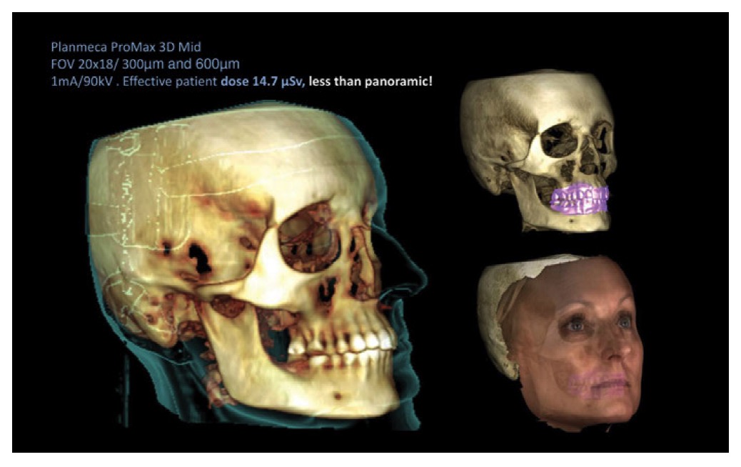
The study examined images taken using the ProMax’s 3D Ultra-Low Dose™ protocol with standard exposures. Images were taken at 24 locations in a 10-year old child and adult phantom, with multiple exposures made for each imaging location. Dosimeters were read 3 times, and dosimeter values were adjusted for sensitivity of dosimeters to affect kV or X-ray source.
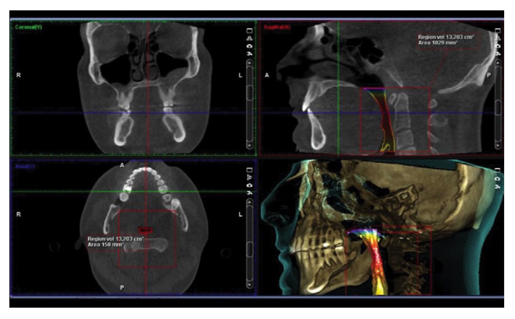
 The researchers found that using the Ultra-Low Dose protocol resulted in an average of 77% reduction in radiation exposure when compared with standard imaging protocols. The researchers also found that while the imaging methods reduced exposure, they found no “statistical reduction in image quality between ULD and standard protocols, suggesting that patient doses can be reduced without loss of diagnostic quality.”
“In my opinion, the ULD images acquired by the Planmeca ProMax in this study meet the standards of the ALARA radiation safety principle,” comments Dr. Jack Fisher, professor of dentistry and orthodontics at Vanderbilt University School of Dentistry. “Why would anyone take a 2D image with this amount of exposure when they can get a 3D image with excellent diagnostic quality at an ultra-low dose of radiation?”
The study was supported, in part, by a grant from the National Institute of Dental and Craniofacial Research (NIDCR). For copies of this study, please contact Planmeca at (855) 245-2908.
*According to “Dosimetry of Orthodontic Diagnostic FOVs Using Low Dose CBCT Protocol.”
This information was provided by Planmeca.
Stay Relevant With Orthodontic Practice US
Join our email list for CE courses and webinars, articles and mores
