Dr. Maria Geisinger addresses the issue of dental office aerosols and possible risks to mitigate.
Dr. Maria L. Geisinger discusses likely modes of transmission for the virus that causes COVID-19

Aerosols created during dental procedures have been suggested as a risk for disease transmission for patients and practitioners in the dental office. The current worldwide pandemic of COVID-19 caused by the transmission of SARS-CoV-2 represents a novel disease and presents unique challenges for our profession moving forward. As we continue to assess transmission rates and modalities, we continue to learn about the risks posed by the airborne transmission of this virus and the risks for dental healthcare professionals and patients. Risk mitigation efforts have been suggested, but such measures should be reasonable and informed by scientific evidence and the known understanding of likely modes of transmission and sub-sequent infectivity.
The CDC has stated that the modes of SARS-CoV-2 transmission are most likely to be via airborne droplets and close, prolonged contact with infectious persons. Furthermore, while the infectivity of asymptomatic carriers is questionable, presymptomatic individuals have been demonstrated to be capable of viral spread. The risk of spread of infection by aerosolized particles may be affected by the type of aerosol generated, aerosol kinetics, pathogen bioload in the aerosol source, and the type of pathogen. Practically, within the dental office, it is important for dental practitioners to be familiar with the following:
- Risks associated with differing modes of transmission, including droplets, aerosols, and fomite surfaces
- Types of aerosols generated by common dental procedures
- The nature, quantity, and sources of microbial load in such aerosols
- The efficacy of current and emerging practices in mitigating aerosol-generated microbial load
Splatter and aerosols: physical and microbial properties
It is well established that airborne infections may be transmitted via droplets, including splatter and aerosols.1 Splatter droplets are defined as those with particle sizes ≥50µm, generally act as a projectile, and are only airborne briefly prior to hitting a surface or falling to earth with gravitational forces. They are spread by close contact (typically within 1 meter) with the host. The particle size of aerosols is <50µm. They may remain airborne for prolonged periods of time, carry viable pathogens, and are capable of being deposited on distant surfaces. It has been demonstrated that droplets >5µm generally remain in the upper respiratory tract while droplets ≤5µm can be inhaled into the lower respiratory tract and those ≤1µm can enter alveoli2,3 (Figure 1).

The interaction and kinetics of droplets in aerosols and splatter are complex. In general, aerosols are always produced in conjunction with splatter, and aerosol droplets may collide with each other, causing them to coalesce and altering their size and physical properties.4 Additionally, larger droplet particles may break down into smaller particles, which may influence the microbial load and particle size.
Droplets, including splatter and aerosols, are routinely generated during physiologic activities such as breathing, talking, coughing, and sneezing. The microbial load created from these activities also differ based upon the type of microorganisms, the infectious course of each disease, and mitigation strategies such as patients wearing surgical masks.5-11 Given these findings, it can be assumed that there is signficant hetero-geneity in the amount, types, and infectivity of droplets produced by individuals in vivo.
The oral cavity contains an extremely diverse microbiome and generally contains a high number of microorganisms. Exogenous infections within or originating from the oral cavity may be influenced by the exposure to and pathogenicity of the infectious organism as well as host susceptibility. Host factors that may influence infectivity include age, immune-inflammatory status, smoking status, and/or concomitant microbial infections.12-15
Aerosols in the dental office
It is well established that many dental procedures produce splatter and aerosols. The highest amounts of aerosols produced during dental procedures are derived from the use of powered scalers, high-speed handpieces, and/or laser use.
Powered scaler use
Sonic, ultrasonic, or piezoelectric devices all produce high levels of aerosol, and the amounts and distance traveled of these droplets is comparable among these devices.16-19 Aerosols produced by ultrasonic instrumentation have been detected as far away as 2 to 11 meters from the treatment site, which could extend throughout dental operatories or offices.20,21 In clinical settings, the levels of aerosols return to preoperative levels within 30 minutes to 2 hours.22,23
High-speed handpiece use
High-speed handpieces can generate droplets, including splatter and aerosols containing blood and other components.20,24-27 The microbial bioload generally correlates with the microbiota present in the tooth being treated26 and the extent of caries in individual patients.28 It has been reported that microbial fallout from restorative procedures can extend up to 1.5 to 2 meters; however, this may be mitigated by the type of evacuation used, and this has not been fully reported.29
Laser instrumentation
Class IV lasers used in dentistry to excise tissue do so through vaporization. This process generates gaseous material often referred to as a smoke plume, which is composed of 95% water.30 The remaining 5% has been reported to contain blood, particulate, and microbial matter. The particle size generated by lasers ranges from 0.1-2µm.30 Although there is no evidence available on disease transmission associated with lasers used in dental operatories, Escherichia coli, Staphylococcus aureus, human papillomavirus, human immunodeficiency virus, and hepatitis B virus have been detected in medical laser plumes.30
Clinical implications
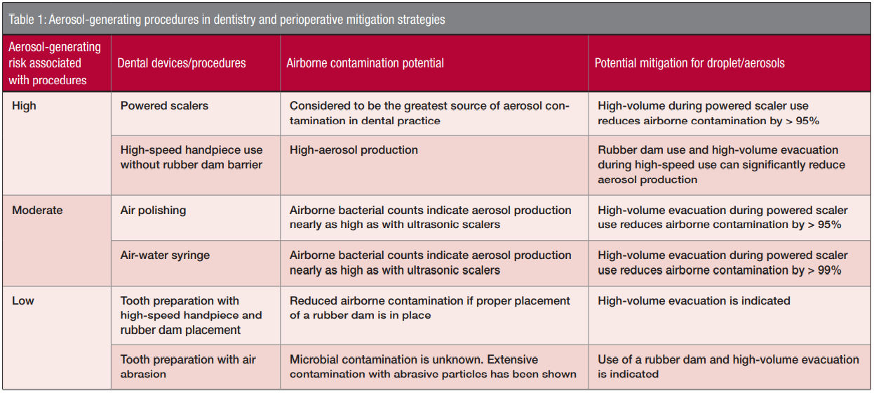
While much focus has been placed on the theoretical risk of disease transmission from dental aerosols, there is limited data identifying the source and infective potential of pathogens in such aerosols. Microbes such as Staphylococcus aureus, beta hemolytic Streptococci, Escherichia coli, spore-forming bacteria, fungi belonging to the genera Cladosporium and Penicillium, and Micrococccus have been identified in dental aerosols.21,31-33 As such, these findings may indicate that microbial sources potentially include saliva, dental water reservoirs, including dental unit water lines (DUWL), or respiratory droplets, with the majority of cultivatable organisms derived from non-patient sources. Water coolant used in conjunction with rotary handpieces and powered scalers has a typical flow rate of 10 to 40 mL per minute34 generally five- to 10-fold greater than unstimulated and stimulated saliva; it can be theorized that significant dilution of salivary or respiratory pathogens occurs in these settings. Because it is expected that SARS-CoV-2 and other airborne pathogens are likely transmitted via human secretions, this dilution may prove to reduce the overall pathogenic bioload and, therefore, infectivity of such aerosols (Table 1).

Bioaerosol mitigation in the dental office
Although there is no evidence implicating dental procedures in the spread of viral particles, the recent COVID-19 epidemic has created an increased awareness of airborne disease transmission and has increased interest in the potential to mitigate aerosol exposure for dental healthcare providers and patients. Strategies to achieve this may include the following:
- Better identifying potentially infectious individuals
- Reducing aerosol bioload
- Barriers that reduce droplet deposition and aspiration for dental healthcare providers
- Reduction of aerosol droplets in room air.19
Strategies to achieve this may include screening prospective patients for common disease symptoms and/or testing prior to invasive procedures, implementing pre-procedural mouth rinses, use of advanced respiratory protections during aerosol-generating procedures, utilization of high-volume evacuation during procedures known to generate dental aerosols, and implementation of advanced technologies to “scrub” the air, including air filtration and/or ultraviolet light decontamination.19,27,35-40
Gaps in our current understanding
The dental profession continues to have unanswered questions about aerosol production in the dental office and its ability to infect dental practitioners, staff, and subsequent patients. Information regarding the bioload of aerosols produced in clinical dental settings and the necessary infective doses of various airborne pathogens are critical to our understanding of risks. Additional research focusing on factors that influence aerosol spread in dental offices, such as airflow and mitigation strategies, is critical. Lastly, epidemiologic evidence of the prevalence of infections in dental healthcare providers and a comparison to populations as a whole may shine a light on highly protective infection control practices that can be implemented to keep practitioners and patients as safe as possible.
Conclusion
In summary, available evidence suggests the following:
- Aerosols are generated by all individuals during many routine activities, including speaking, eating, and breathing.
- The bioload in aerosols correlates with disease severity for respiratory diseases.
- Aerosols are also created during most dental procedures. The dental procedures associated with the highest levels of aerosols are powered scalers, high-speed handpieces, air-water syringes, and air polishers.
- Most current evidence suggests that dental water reservoirs are the primary source of pathogens in these aerosols, rather than saliva or respiratory secretions.
- Several methods are effective in mitigating the production of dental aerosols and in reducing bioload. Chief among these are the use of high-volume evacuators and pre-procedural mouth rinsing, but effectiveness may vary based upon implementation within dental practices.
- Similarly, several barrier techniques are effective in protecting the occupants of the dental operatory from direct and indirect aerosol exposure. These include commonly used PPE such as surgical masks/respirators, face shields, fluid impermeable gowns, and gloves.
- No evidence exists to suggest that dental healthcare professionals are at a higher risk of airborne viral disease transmission than the general population, and emerging evidence suggests that the risk may be lower during the delivery of dental care than in other healthcare settings.
- Nonclinical areas within the dental office and/or community exposure of dental personnel may pose a significant risk within the dental office, and adherence to public health guidelines is critical to limit spread of airborne illness.
Here is one way to reduce dental office aerosols in the orthodontic office. https://orthopracticeus.com/industry-news/asi-dental-shares-how-to-reduce-aerosols-in-an-open-bay-orthodontic-design/
- Keene CH. Airborne Contagion and Air Hygiene. William Firth Wells. J Sch Health. 1955;25:249-249.
- Annex C—Respiratory Droplets. In: Atkinson J, Chartier Y, Pessoa-Silva CL, et al., eds. Infection Control in Health-Care Settings. WHO Press, World Health Organization. Geneva, Switzerland; 2009. https://www.who.int/water_sanitation_health/publications/natural_ventilation.pdf. Accessed July 6, 2020.
- Micik RE, Miller RL, Mazzarella MA, Ryge G. Studies on dental aerobiology, I: bacterial aerosols generated during dental procedures. J Dent Res. 1969;48(1):49-56.
- Hinds WC. Aerosol Technology: Properties, Behavior, and Measurement of Airborne Particles. 2nd ed. Hoboken, NJ: John Wiley & Sons (Wiley-Interscience); 1999.
- Zheng Y, Chen H, Yao M, Li X. Bacterial pathogens were detected from human exhaled breath using a novel protocol. J Aerosol Sci. 2018;117:224-234.
- Knibbs LD, Johnson GR, Kidd TJ, et al. Viability of Pseudomonas aeruginosa in cough aerosols generated by persons with cystic fibrosis. 2014;69(8):740-745.
- Hatagishi E, Okamoto M, Ohmiya S, et al. Establishment and clinical applications of a portable system for capturing influenza viruses released through coughing. PLoS One. 2014;9(8):e103560.
- Yan J, Grantham M, Pantelic J, et al. Infectious virus in exhaled breath of symptomatic seasonal influenza cases from a college community. Proc Natl Acad Sci USA. 2018;115(5):1081-1086.
- Tang JW, et al. Absence of detectable influenza RNA transmitted via aerosol during various human respiratory activities — experiments from Singapore and Hong Kong. PLoS One. 2014;9;e107338. doi:10.1371/journal.pone.0107338
- Milton DK, Fabian MP, Cowling BJ, Grantham ML, McDevitt JJ. Influenza virus aerosols in human exhaled breath: particle size, culturability, and effect of surgical masks. PLoS Pathog. 2013;9(3).
- Wood ME, Stockwell RE, Johnson GR, et al. Face Masks and Cough Etiquette Reduce the Cough Aerosol Concentration of Pseudomonas aeruginosa in People with Cystic Fibrosis. Am J Respir Crit Care Med. 2018;197(3):348-355.
- To KKW, Yip CCY, Lai CYW, et al. Saliva as a diagnostic specimen for testing respiratory virus by a point-of-care molecular assay: a diagnostic validity study. Clin Microbiol Infect. 2019;25(3):372-378.
- Kim YG, Yun SG, Kim MY, et al. Comparison Between Saliva and Nasopharyngeal Swab Specimens for Detection of Respiratory Viruses by Multiplex Reverse Transcription-PCR. J Clin Microbiol. 2017;55:226-233.
- Tada A, Shiiba M, Yokoe H, Hanada, Tanzawa H. Relationship between oral motor dysfunction and oral bacteria in bedridden elderly. Oral Surg Oral Med Oral Pathol Oral Radiol Endod. 2004;98(2):184-188.
- Tada A, Hanada N, Tanzawa, H. The relation between tube feeding and Pseudomonas aeruginosa detection in the oral cavity. J Gerontol A Biol Sci Med Sci. 2002;57(10):M71-M72.
- Gross KB, Overman, PR, Cobb C, Brockmann S. Aerosol generation by two ultrasonic scalers and one sonic scaler. A comparative study. J Dent Hyg. 1992;66(7):314-318.
- Rivera-Hidalgo F, Barnes JB, Harrel SK. Aerosol and splatter production by focused spray and standard ultrasonic inserts. J Periodontol. 1999;70(5):473-477.
- Graetz C, Plaumann A, Jule Bielfeldt J, et al. Efficacy versus health risks: An in vitro evaluation of power-driven scalers. J Indian Soc Periodontol. 2015;19(1):18-24.
- Harrel SK, Barnes JB, Rivera-Hidalgo F. Aerosol and splatter contamination from the operative site during ultrasonic scaling. J Am Dent Assoc. 1998;129(9):1241-1249.
- Grenier D. Quantitative analysis of bacterial aerosols in two different dental clinic environments. Appl Environ Microbiol. 1995;61(8):3165-3168.
- Singh A, Shiva Manjunath RG, Singla D, et al. Aerosol, a health hazard during ultrasonic scaling: A clinico-microbiological study. Indian J Dent Res. 2016;27(2):160-162.
- Dutil S, Meriaux A, de Latremoille M-C, et al. Measurement of airborne bacteria and endotoxin generated during dental cleaning. J Occup Environ Hyg. 2009;6(2):121-130.
- Veena HR, Mahantesha S, Joseph PA, Patil SR, Patil SH. Dissemination of aerosol and splatter during ultrasonic scaling: a pilot study. J Infect Public Health. 2015;8(3): 260-265.
- Bennett AM, Fulford MR, Walker JT, et al. Microbial aerosols in general dental practice. Br Dent J. 2000;189(12):664-667.
- Osorio R, Toledano M, Liébana J, Rosales JI, Lozano JA. Environmental microbial contamination. Pilot study in a dental surgery. Int Dent J. 1995;45(6):352-357.
- Bentley CD, Burkhart NW, Crawford JJ. Evaluating spatter and aerosol contamination during dental procedures. J Am Dent Assoc. 1994;125(5):579-584.
- Yamada H, Ishihama K, Yasuda K, et al. Aerial dispersal of blood-contaminated aerosols during dental procedures. Quintessence Int. 2011;42(5);399-405.
- Serban D, Banu A, Serban C. Tuta-Sas I, Vlaicu B. Predictors of quantitative microbiological analysis of spatter and aerosolization during scaling. Rev Med Chir Soc Med Nat Iasi. 2013;117:503-508.
- Rautemaa R, Nordberg A, Wuolijoki-Saaristo K, Meurman JH. Bacterial aerosols in dental practice — a potential hospital infection problem? J Hosp Infect. 2006;64(1):76-81.
- Bargman H. Laser-generated Airborne Contaminants. J Clin Aesthet Dermatol. 2011;4(2):56-57.
- Hallier C, Williams DW, Potts AJC, Lewis MAO. A pilot study of bioaerosol reduction using an air cleaning system during dental procedures. Br Dent J. 2010;209(8):E14,
- Teanpaisan R, Taeporamaysamai M, Rattanachone P, Poldoung N, Srisintorn S. The usefulness of the modified extra-oral vacuum aspirator (EOVA) from household vacuum cleaner in reducing bacteria in dental aerosols. Int Dent J. 2001;51(6):413-416.
- Kobza J, Pastuszka JS, Bragoszewska E. Do exposures to aerosols pose a risk to dental professionals? Occup Med (Lond). 2018;68(7):454-458.
- Lea SC, Landini G, Walmsley AD. Thermal imaging of ultrasonic scaler tips during tooth instrumentation. J Clin Periodontol. 2004;31(5):370-375.
- Jacks ME. A laboratory comparison of evacuation devices on aerosol reduction. J Dent Hyg. 2002;76(3):202-206.
- Yadav N, Agrawal B, Maheshwari C. Role of high-efficiency particulate arrestor filters in control of air borne infections in dental clinics. SRM J Res Dent Sci. 2015;6:240-242.
- American Society for Healthcare Engineering. ASHE™ website. Air filtration. https://www.ashe.org/compliance/ec_02_05_01/01/airfiltration. Accessed July 5, 2020.
- Chen C, Zhao B, Cui W, et al. The effectiveness of an air cleaner in controlling droplet/aerosol particle dispersion emitted from a patient’s mouth in the indoor environment of dental clinics. J R Soc Interface. 2010;7(48):1105-1118.
- Alexander DD, Bailey WH, Perez V, Mitchell ME, Su S. Air ions and respiratory function outcomes: a comprehensive review. J Negat Results Biomed. 2013;12:14.
- Lindblad M, Tano E, Lindahl C, Huss F. Ultraviolet-C decontamination of a hospital room: Amount of UV light needed. 2020;46(4)842-849.
Stay Relevant With Orthodontic Practice US
Join our email list for CE courses and webinars, articles and mores

 Maria L. Geisinger, DDS, MS, is a Professor and Director of Advanced Education in Periodontology in the Department of Periodontology at the University of Alabama at Birmingham (UAB) School of Dentistry. Dr. Geisinger received her BS in Biology from Duke University, her DDS from Columbia University School of Dental Medicine, and her MS and Certificate in Periodontology and Implantology from The University of Texas Health Science Center at San Antonio. Dr. Geisinger is a Diplomate of the American Board of Periodontology. She has served as the President of the American Academy of Periodontology Foundation and on multiple nationally and regionally organized dentistry committees. She currently serves as Chair of the ADA’s Council on Scientific Affairs and is a member of the AAP’s Board of Trustees. She has authored over 45 peer-reviewed publications. Her research interests include periodontal and systemic disease interaction, implant dentistry in the periodontally compromised dentition, and novel treatment strategies for oral soft and hard tissue growth. Dr. Geisinger lectures nationally and internationally on topics in periodontology and oral healthcare.
