Drs. Jin-Young Choi, Jun-Ho Park, Hong-Chul Yoon, and Seong-Hun Kim discuss an innovative method using Digital Quantitative Light-Induced Fluorescence (QLF) in detecting clinical issues in orthodontic patients
Orthodontic treatment is a complex procedure that necessitates consideration of not only dental esthetics but also masticatory function and overall oral health. Patients utilizing fixed orthodontic appliances such as brackets and bands frequently encounter challenges in maintaining optimal oral hygiene. This difficulty leads to an increased risk of periodontal disease and dental caries, in addition to the formation of microscopic cracks that are not readily detectable with the naked eye. In practice, relying solely on patient self-care during orthodontic therapy often proves insufficient, as microscopic lesions can readily develop in blind spots such as around brackets, interproximal surfaces, and the lingual aspects of teeth. Consequently, concern regarding the development of caries, periodontal disease, and the potential weakening of teeth during orthodontic treatment, as well as uncertainty about the appropriate timing for intervention or treatment, are common among patients, guardians, and dental practitioners alike.
Traditionally, the evaluation of dental and periodontal health has depended primarily on visual inspection or, when deemed necessary, radiographic imaging techniques such as panoramic, bitewing, and periapical radiographs. Although these methods are sensitive to advanced lesions, they present significant limitations in detecting early-stage pathologies, including initial demineralization, white spot lesions, micro-cracks, and incipient carious lesions. Furthermore, repeated radiographic examinations pose the risk of cumulative radiation exposure, are subject to anatomical blind spots, and cannot reflect the real-time progression and activity of lesions.
An innovative diagnostic method that addresses these clinical concerns through digital data is based on Quantitative Light-induced Fluorescence (QLF) (Q-ray system, AIOBIO, Seoul, South Korea). QLF utilizes blue visible light at a wavelength of 405 nm to illuminate the tooth surface: healthy regions of the tooth exhibit green fluorescence, while regions with demineralization (such as early carious lesions), micro-cracks, or accumulations of bacterial metabolites display either loss of fluorescence or red fluorescence due to scattered light. These subtle fluorescence patterns are quantitatively represented as ∆F (indicating mineral loss) and ∆R (representing porphyrin-based bacterial metabolites), providing numerical data for objective assessment.
Based on this principle, this technology enables real-time and repeated detection of otherwise hidden lesions during orthodontic treatment. For example, white spot lesions that appear around brackets — indicators of initial demineralization which may be missed by visual inspection or clinical experience alone — can be detected at an early stage, as the ∆F value exceeds a defined threshold. Similarly, interproximal caries, which are traditionally undetectable without bitewing radiographs, can be precisely identified by Q-ray even when mineral loss is less than 6%. Most importantly, micro-cracks in teeth, which are almost impossible to detect visually or radiographically during orthodontic treatment, can be visualized, with the location, depth, and bacterial activity of the lesion clearly displayed via ∆F, ∆R values, and fluorescence imaging — crucial information for prognosis and treatment planning.
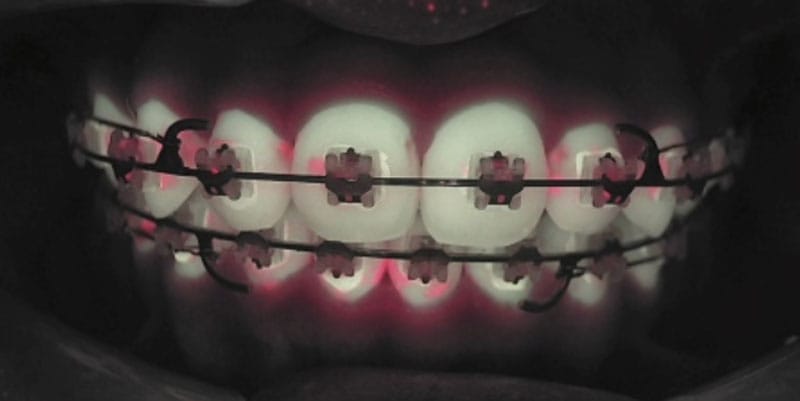
The clinical potential of this type of technology is substantiated by objective data. At Kyung Hee University Dental Hospital, an analysis of over 150 subjects and more than 300 teeth revealed that more than half of early carious lesions, micro-cracks, and subtle interproximal defects, which could only be detected using Q-ray, were missed by conventional diagnostic techniques. The benefits extend beyond caries and cracks; one of its key strengths lies in enhancing communication between patients and practitioners. All examination outcomes are stored as photographic images, color-mapped graphics, and quantitative data, enabling visualization of lesion changes over time — from the initiation of orthodontic treatment, through interim monitoring, to post-removal of appliances. As a result, patients are not merely given fleeting verbal explanations but are presented with objective data on the status of their teeth, which strongly motivates them toward improved oral hygiene and increases their engagement with the treatment process. The images above present Q-ray scans from patients with labial and lingual bracket attachments. Presenting such imagery to patients not only underscores the importance of oral hygiene, increasing the educational effect, but also provides objective evidence that may protect clinicians in the event of dental caries or other complications arising during orthodontic treatment.
The extensive clinical potential of Q-ray technology is founded upon its originality, reliability, and ease of use. The QLF device developed by AIOBIO has successfully obtained over 20 international patents and has been certified by major global regulatory bodies, including CE marking, FDA clearance, and approval by the Korean Ministry of Food and Drug Safety. Integrated with the advanced QA2 analytical software, the system facilitates seamless processes encompassing region of interest (ROI) designation, quantitative calculation of ∆F/∆R values, and comprehensive patient-specific visualization data management. Furthermore, the absence of ionizing radiation exposure and the brief examination time render this technology particularly suitable and convenient for diverse patient populations, including children, the elderly, pregnant women, and individuals requiring frequent, periodic monitoring.
Clinical feedback regarding the implementation of the device within the Biocreative Orthodontics Strategy Center at Kyung Hee University Dental Hospital has been notably positive. Clinicians have reported a marked reduction in overlooked early carious lesions and cracks during orthodontic treatment. Moreover, the reliance on conventional radiographic methods has diminished, enabling practitioners to provide patients with clear, visualization-based explanations grounded in objective quantitative data. Patient and caregiver satisfaction has significantly increased due to the availability of visual imagery and numerical assessments, which has correspondingly led to a substantial decrease in complaints related to dental caries, cracks, and subtle interproximal lesions occurring throughout orthodontic therapy.
Q-ray–based diagnostics in orthodontic patients sets the stage for a new era of data-driven, visualization-based oral health management extending beyond orthodontics to the broader field of dentistry.
References
- Oh SH, Lee SR, Choi JY, Choi YS, Kim SH, Yoon HC, Nelson G. Detection of Dental Caries and Cracks with Quantitative Light-Induced Fluorescence in Comparison to Radiographic and Visual Examination: A Retrospective Case Study. Sensors (Basel). 2021 Mar 3;21(5):1741. doi: 10.3390/s21051741.
- Oh SH, Choi JY, Kim SH. Evaluation of dental caries detection with quantitative light-induced fluorescence in comparison to different field of view devices. Sci Rep. 2022 Apr 12;12(1):6139. doi: 10.1038/s41598-022-10126-x.
Stay Relevant With Orthodontic Practice US
Join our email list for CE courses and webinars, articles and mores
